Open Access, Peer-reviewed
eISSN 2093-9752

Open Access, Peer-reviewed
eISSN 2093-9752
Min Ju Shin
YoungJin Moon
Sang Ki Lee
Hwi-Yeol Yun
JuWon Song
Zheng Dong
Du-Bin Im
JiaHao Xu
XuanRu Wang
http://dx.doi.org/10.5103/KJAB.2024.34.4.204 Epub 2024 December 22
Abstract
Objective: The aim of this study was to investigate the impact of cognitive recognition and avoidance movement performance in the moments before a fall on fall risk among the elderly.
Method: 8 men (age: 73.1 ± 4.1 years, height: 166.3 ± 4.4 cm, weight: 66.0 ± 8.0 kg) and 15 women (age: 70.8 ± 3.6 years, height: 155.5 ± 5.1 cm, weight: 58.2 ± 8.4 kg) total 23 elderly participated in this study. Participants were evaluated for fall risk using the Berg Balance Scale (BBS) and Time Up and Go (TUG) test. Electromyography (EMG) and electroencephalography (EEG) were recorded during a fall avoidance task to assess muscle activation and cognitive responses.
Results: The study revealed a significant negative correlation between age and BBS scores, and a positive correlation between age and TUG completion times, indicating that older participants had reduced balance and slower mobility. Additionally, an increase in antagonist muscle activity (BF_av) was positively correlated with longer TUG times, suggesting that elevated antagonist co-contraction may hinder efficient movement control during fall avoidance. EEG analysis showed no significant correlations between fall risk scores (BBS and TUG) and ERP components, but specific ERP measures indicated delayed cognitive processing times (P1_LT) were linked to decreased primary muscle activation (GA_av) and increased antagonist activity (TA_av), suggesting that slower cognitive responses may reduce the effectiveness of rapid avoidance movements. These findings underscore the interconnected decline in balance, muscle response, and cognitive processing as factors contributing to increased fall risk in the elderly.
Conclusion: Declines in balance, mobility, and cognitive processing are associated with an increased risk of falls in the elderly, highlighting the need for targeted interventions to improve fall avoidance.
Keywords
Elderly Fall risk Electromyography Electroencephalography Co-contraction
한국은 고령화 속도가 전 세계 국가 중 가장 빠른 나라이며, 2050년에는 고령 인구의 비율이 세계에서 두 번째로 높은 나라가 될 것이라 전망하고 있다(Jang, Lee & Lee, 2019). 저조한 출산율이 지속되면서 노인 인구의 비율이 늘어나게 되었고, 급속한 고령화로 인하여 2021년 65세 이상 고령 인구가 전체 인구의 16.5%였으며, 2025년에는 20.3% 그리고 2060년에는 43.9%가 될 것이라 예상하고 있다(Statics Korea, 2021). 노인 인구의 비율이 증가하면서 노인들의 안전과 관련된 사고 발생 빈도 또한 증가하고 있는데(Seong, Seo, Seol, Shin & Yang, 2020), 그 중 낙상 사고는 노인의 주요 사망 및 부상의 원인 중 하나(Jamebozorgi, Kavoosi, Shafiee, Kahlaee & Raei, 2013). 65세 이상을 기준으로 연령이 높아질수록 낙상의 위험은 더욱 커진다(Zhu & Lin, 2012).
낙상은 예상하지 못 한 상황에서 스스로 신체의 움직임을 제어하지 못할 때 발생하며, 특히 노인에서 발생률이 높다. 2020년 보건복지부에서 실시한 노인실태조사에 따르면 노인 인구 7.2%가 1년에 한 번 이상 낙상을 경험하였으며, 낙상 후 내원 비율은 72.5%라 밝힌 바 있다. 이처럼 노인에게 낙상이 위험한 것으로 인식되어 오고 있지만, 노인 낙상 인구의 꾸준한 증가는 여전히 문제는 해결되지 않고 있다는 증거이다(Seong et al., 2020).
노인 낙상에 미치는 요인들은 많이 보고되고 있으나, 요인 간의 상관관계 및 인과관계 그리고 어떠한 요인들의 상호 복합작용이 이루어지는 것인가에 대한 정보는 많이 부족한 실정이다(Choi, Lim, Kim & Lee 2021). 노인 낙상의 주된 원인은 근육량과 근력의 감소, 신체 균형 능력의 저하와 같은 움직임 제어 능력의 감소, 인지기능의 저하 등으로 보고된다(Deschenes, 2004; Bruce-Keller et al., 2012; Casas Herrero, Martínez Velilla & Alonso Renedo, 2011). 신체의 균형은 고유수용성감각, 시각, 체성감각 그리고 전정계의 상호작용을 통해 유지된다(Shumway-Cook & Horak, 1986). 연령이 증가함에 따라 신경계와 감각계의 약화로 인하여 인지기능의 저하를 초래한다(Sturnieks, George & Lord, 2008). 균형감각 및 인지기능의 저하는 노인의 낙상 증가와 관련이 있으며, 이는 균형 제어가 단순한 관절의 제어가 아닌 신체적 능력과 인지기능의 결합임을 시사한다. 낙상과 부상을 방지하기 위해서는 위험한 상황에 대한 인식 후 적절한 대응이 필요하다(Stelmach & Worringham, 1985).
사건 관련 전위(evenet related potentials, ERP)는 빛과 소리 또는 특정 자극(event)에 관련하여 인지에 대한 신경 반응을 보이는 뇌에서 기록된 전기 신호이다. 특히 주의력, 기억력, 언어 처리 및 의사 결정과 같은 인지기능을 이해하는데 중요한 뇌의 프로세스를 제공한다(Bressler & Ding, 2006). 노화가 진행됨에 따라 시각적 자극에 대한 반응시간(reaction time, RT)이 증가하였으며(Woods, Wyma, Yund, Herron & Reed, 2015), 이는 낙상의 위험 요인임을 암시한다(Lord & Fitzpatrick, 2001). 시각 유발 전위(Visual evoked potnstials, VEP)는 시각적 자극에 대한 후두엽 피질의 반응을 측정하며, 시각적 정보 처리 능력을 평가하는데 사용된다(Sherman, 1979). VEP에서 가장 잘 알려진 파형은 P100(혹은 P1)으로 자극이 제시된 후 약 100 ms 부근에서 발현되는 양전위(positive potentials)이다(Drislane, 2007). 노인 치매 환자 그룹이 동일 연령 노인 그룹에 비해 P100의 잠복기(latency) 더 긴 것을 발견하였다(Pollock et al., 1989). 이는 P100의 잠복기가 시각적 기능뿐 아니라 인지기능의 지표임을 의미한다(Covey et al., 2021). ERP를 통한 각 구성 요소의 분석은 인지적 정보 처리 단계를 조사하는 유익한 방법임을 알 수 있다(Ouyang, Herzmann, Zhou & Sommer, 2011; Sokhadze et al., 2017).
관절은 주동근(agonist)과 그에 대항하는 길항근(antagonist)의 상호작용으로 안정적으로 움직인다(Kim & Hwang, 2018). 연령에 따라 근활성도의 차이가 나타나는데 노화가 진행됨에 따라 노인들은 안정성을 확보하기 위해 서로 대항하는 두 근육군을 동시수축(co-contraction) 하는 전략을 세운다(Craig, Goble & Doumas, 2016; Iwamoto, Takahashi & Shinkoda, 2017). 가만히 서 있는 상황에서 노인들의 동시수축은 정적인 자세를 유지하는 데에 도움이 되지만(Lord, Clark & Webster, 1991), 보행과 같은 동적 균형 제어가 필요한 상황에서는 불안정성을 유발한다(Iwamoto et al., 2017). 또한 근육의 피로를 유발하고, 예상치 못한 장애물이나 상황에 대한 신체적 대처 능력을 감소시켜 낙상의 위험을 증가시킬 수 있다(Nelson-Wong et al., 2012).
낙상은 예상하지 못한 상황일수록 그리고 상황에 대한 인지 능력과 신체적 제어 능력이 감소할수록 위험이 증가하며, 이러한 능력들은 노화가 진행됨에 따라 감소한다. 따라서 본 연구는 노인을 대상으로 낙상 직전 상황에 대한 인지와 그에 따른 낙상 회피 동작 수행 능력이 낙상 위험에 미치는 영향을 규명하고자 한다.
1. 연구 대상자
본 연구의 대상자는 65세 이상 노인 남자 8명(나이: 73.1±4.1 yrs, 키: 166.3±4.4 cm, 몸무게: 66.0±8.0 ㎏), 여자 15명(나이: 70.8±3.6 yrs, 키: 155.5±5.1 cm, 몸무게: 58.2±8.4 ㎏) 총 23명을 대상으로 하였다. 연구의 목적과 내용을 설명하고 자발적인 참여 동의를 얻어 연구를 진행하였다. 본 연구는 충남대학교 연구윤리위원회에서 심의하여 승인을 받고 연구 윤리에 따라 연구를 진행하였다(202310-SB-179-01).
2. 실험 방법
본 연구에 참여한 모든 피험자는 낙상 위험 평가를 위해 버그 균형 척도(Berg Balance Scale, BBS)와 Time Up and Go (TUG) 검사를 진행하였다. BBS와 TUG 검사는 점수가 낮을수록 낙상의 위험이 증가한다는 결과를 통해 낙상의 위험을 효과적으로 예측하는데 타당한 도구임을 확인하였고(Shumway-Cook, Brauer & Woollacott, 2000; Lajoie & Gallagher, 2004), 일관된 결과를 제공하는 신뢰도가 높은 도구임을 보고하였다(Conradsson et al., 2007; Steffen, Hacker & Mollinger, 2002). BBS는 14개 항목으로 구성되어 있으며, 앉은 상태에서의 균형, 제자리 선 상태에서의 정적 균형, 동적 균형을 포함하고 있다. 각 항목 당 최대 점수는 4점으로 45점 이하일 경우 낙상 위험이 높은 것으로 평가한다. TUG 검사는 의자에 앉아서 시작하여 '시작' 신호와 동시에 의자에 일어나 3 m를 걷고 반환점을 돌아와 다시 의자에 '앉기' 까지의 시간(초)를 측정하였다. 이때 총 왕복 시간이 12초 이상일 경우 낙상 위험도가 높은 것으로 평가한다.
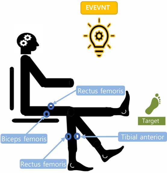
ERP와 하지 근육의 공동 수축을 평가하기 위해 뇌파 측정은 BIOS-ST (BioBrain, Korea)와 근전도 측정은 Wave EMG (Cometa System, Italy)을 이용하여 trigger를 사용하여 동시 측정하였다. 실험 대상자는 의자에 앉아 정면을 주시하고 외부 자극(빛)이 시작되면 사전에 지시한 동작을 수행하였다. 사전에 지시한 동작은 무릎을 펴고(extension) 발목을 저측굴곡(plantar flexion)하여 목표지점을 누르는 것이다. 본 실험에서 외부 자극은 전방 낙상 상황을 의미하고, 사전에 지시한 동작은 전방 낙상 시 회피를 위한 대응 동작으로 정의하였다(Madigan & Lloyd, 2005). 실험 동작은 약 40회 반복 수행하였으며, <Figure 1>과 같이 실험을 설계하였다.

3. 데이터 처리
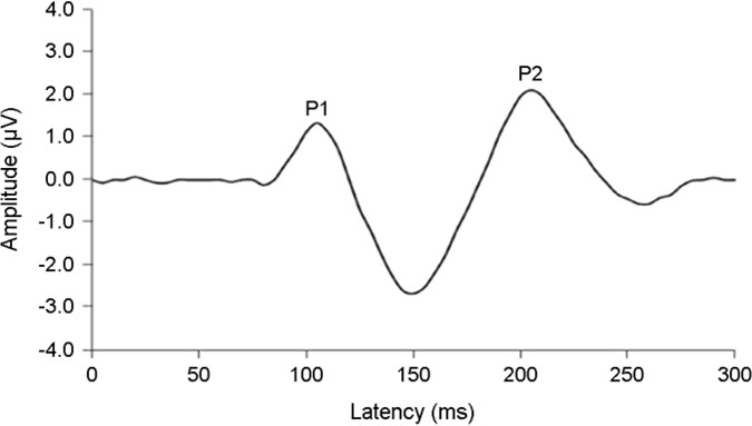
본 연구의 모든 데이터는 실험 동작 40회 중 시작과 끝 각각 10회 동작을 제외한 20회 반복 수행한 데이터를 사용하였다. 무선 근전도 EMG를 사용하여 넙다리곧은근(RF), 넙다리두갈래근(BF), 장딴지근(GA), 앞정강근(TA)의 RMS 값을 통해 근활성도 값을 산출하였으며, 뇌파는 시각적 자극에 대한 반응이 가장 강하게 나타나는 후두부 위치 O1, O2의 P1과 P2의 평균 진폭(amplitude, A)과 잠복기(latency, LT)를 분석하였다(Figure 2 참고).
1) 동시수축 비율(Co-Contraction Ratio)
다음과 같은 관계식에서 주동근과 길항근의 동시수축 비율이 산출된다.
4. 통계 분석
본 연구의 통계 처리는 SPSS 26.0을 사용하여 낙상 위험 평가 점수와 하지 근육의 공동 활성과 평균 활성 그리고 ERP 간의 상관관계 분석을 진행하였다. 데이터는 Shapiro-Wilk test를 사용하여 데이터의 정규 분포 확인 후 Pearson's correl- ation을 사용하여 다중상관분석(multiple correlation analysis)을 진행하였다. 통계적 유의수준 α=.05로 설정하였다.
1. 낙상 위험도와 근육, ERP의 상관관계 결과
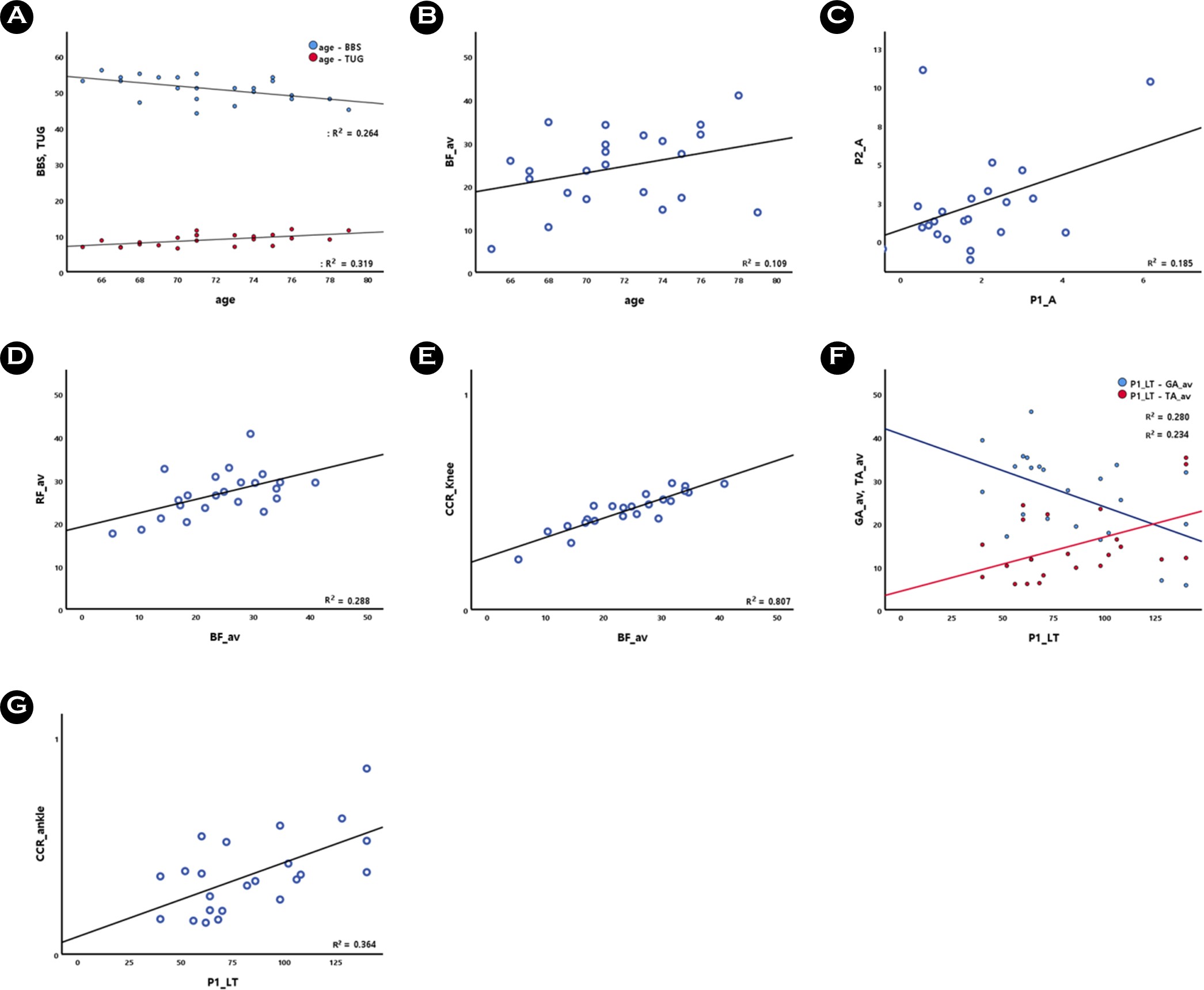
BBS의 점수는 연령(age)과 -.514*로 유의한 음의 상관관계를 보였다(p<.05). TUG 소요시간은 연령(age)과 .565**로 유의한 양의 상관관계를 보였고(p<.01), BF_av와 .440*으로 유의한 양의 상관관계를 보였다(p<.05). 그러나 낙상 위험도(BBS와 TUG)와 ERP 간의 유의한 상관관계는 나타나지 않았다(Table 1, Figure 3).
|
|
BBS |
|
TUG |
|||
|
r |
p |
r |
p |
|||
|
Muscle |
Age |
-.514* |
0.012 |
|
.565** |
0.005 |
|
RF_av |
-0.148 |
0.500 |
|
0.322 |
0.134 |
|
|
BF_av |
-0.118 |
0.592 |
|
.440* |
0.036 |
|
|
CCR_knee |
-0.008 |
0.973 |
|
0.357 |
0.095 |
|
|
GA_av |
0.080 |
0.716 |
|
-0.182 |
0.407 |
|
|
TA_av |
0.217 |
0.319 |
|
0.238 |
0.275 |
|
|
CCR_ankle |
0.113 |
0.609 |
|
0.254 |
0.243 |
|
|
ERP |
P1_A |
-0.033 |
0.883 |
|
0.091 |
0.680 |
|
P1_LT |
0.126 |
0.566 |
|
-0.026 |
0.908 |
|
|
N1_A |
0.151 |
0.490 |
|
-0.190 |
0.384 |
|
|
N1_LT |
-0.263 |
0.226 |
|
0.076 |
0.732 |
|
|
P2_A |
-0.020 |
0.927 |
|
0.197 |
0.367 |
|
|
P2_LT |
-0.003 |
0.991 |
|
0.111 |
0.615 |
|
|
NOTE. RF_av: Average
activity of the rectus femoris, BF_av: Average activity of the biceps femoris,
CCR_knee: Co-contraction ratio of the knee, GA_av: Average activity of the
gastrocnemius, TA_av: Average activity of the tibialis anterior, CCR_ankle: Co-contraction
ratio of the ankle, P1_A: Amplitude of P1, P1_LT: Latency of P1, N1_A: Amplitude
of N1, N1_LT: Latency of N1, P2_A: Amplitude of P2, P2_LT: Latencey of P2 significant at *p<.05,
**p<.01 |
||||||
2. 인지기능의 상관관계 결과
P1_A는 P2_A와 .430*로 유의한 양의 상관관계를 보였지만(p<.05), 나머지 변인 간의 상관관계는 보이지 않았다(Table 2, Figure 3).
|
|
P1_A |
P1_LT |
N1_A |
N1_LT |
P2_A |
P2_LT |
|
|
P1_A |
1 |
|
|
|
|
|
|
|
P1_LT |
0.202 |
1 |
|
|
|
|
|
|
N1_A |
-0.333 |
0.190 |
1 |
|
|
|
|
|
N1_LT |
0.289 |
0.272 |
0.195 |
1 |
|
|
|
|
P2_A |
.430* |
0.236 |
-0.354 |
0.101 |
1 |
|
|
|
P2_LT |
0.252 |
0.154 |
-0.329 |
-0.043 |
0.128 |
1 |
|
|
NOTE. P1_A: Amplitude of P1, P1_LT: Latency of P1,
N1_A: Amplitude of N1, N1_LT: Latency of N1, P2_A: Amplitude of P2, P2_LT: Latencey
of P2 significant at *p<.05 |
|||||||
3. 움직임 제어의 상관관계 결과
RF_av는 BF와 .537**로 유의한 양의 상관관계를 보였고, BF_av는 CCR_knee와 .899**로 유의한 양의 상관관계를 보였다(p<.01). 또한 CCR_ankle은 GA_av와 -.807**로 유의한 음의 상관관계를 보였으며, TA_av와 .808**로 유의한 양의 상관관계를 보였다(p<.01, Table 3, Figure 2).
|
|
RF_av |
BF_av |
CCR_knee |
GA_av |
TA_av |
CCR_ankle |
|
|
RF_av |
1 |
|
|
|
|
|
|
|
BF_av |
.537** |
1 |
|
|
|
|
|
|
CCR_knee |
0.198 |
.898** |
1 |
|
|
|
|
|
GA_av |
0.152 |
-0.100 |
-0.182 |
1 |
|
|
|
|
TA_av |
0.209 |
0.411 |
0.400 |
-0.354 |
1 |
|
|
|
CCR_ankle |
0.065 |
0.277 |
0.320 |
-.807** |
.808** |
1 |
|
|
NOTE. RF_av: Average
activity of the rectus femoris, BF_av: Average activity of the biceps
femoris, CCR_knee: Co-contraction ratio of the knee, GA_av: Average activity
of the gastrocnemius, TA_av: Average activity of the tibialis anterior, CCR_ankle:
Co-contraction ratio of the ankle significant at **p<.01 |
|||||||

4. 인지기능과 움직임 제어의 상관관계 결과
P1_LT는 GA_av와 -.529**로 유의한 양의 상관관계를 보였고(p<.01), TA_av와 .484*로 유의한 양의 상관관계를 보였다(p<.05). 또한 P1_LT는 CCR_ankle과 .604**로 유의한 양의 상관관계를 나타냈다(p<.01, Table 4, Figure 3, 참조).
|
|
P1_A |
P1_LT |
N1_A |
N1_LT |
P2_A |
P2_LT |
|
|
RF_av |
-0.007 |
0.065 |
-0.214 |
-0.097 |
0.216 |
0.353 |
|
|
BF_av |
-0.374 |
0.031 |
0.038 |
-0.041 |
0.072 |
0.089 |
|
|
CCR_knee |
-0.365 |
0.032 |
0.188 |
-0.006 |
0.041 |
0.049 |
|
|
GA_av |
0.007 |
-.529** |
-0.167 |
-0.099 |
0.017 |
-0.149 |
|
|
TA_av |
-0.197 |
.484* |
0.113 |
0.023 |
0.226 |
0.077 |
|
|
CCR_ankle |
-0.094 |
.604** |
0.169 |
0.062 |
0.181 |
0.099 |
|
|
NOTE. RF_av: Average
activity of the rectus femoris, BF_av: Average activity of the biceps
femoris, CCR_knee: Co-contraction ratio of the knee, GA_av: Average activity
of the gastrocnemius, TA_av: Average activity of the tibialis anterior, CCR_ankle:
Co-contraction ratio of the ankle, P1_A: Amplitude of P1, P1_LT: Latency of P1,
N1_A: Amplitude of N1, N1_LT: Latency of N1, P2_A: Amplitude of P2, P2_LT: Latencey
of P2 significant at *p<.05, **p<.01 |
|||||||

본 연구에서는 노인들의 낙상 위험도와 낙상 유사 상황에 대한 인지 그리고 낙상 회피 동작 수행 시 평균 근활성 및 동시 수축의 상관관계에 대해 조사하였다. 연령과 신체의 균형을 평가하는 BBS의 점수는 음의 상관관계를 가지고 있으며, 이동성을 평가하는 TUG의 소요시간은 양의 상관관계를 보여주고 있다. 이는 연령이 증가함에 따라 자세의 안정성과 이동성이 감소한다는 이전 연구와 일치하며(Bao et al., 2018; Trombetti et al., 2016), BBS와 TUG는 낙상의 위험을 예측하는 주요 도구(Ambrose, Paul & Hausdorff, 2013)임이 또다시 확인되었다. 또한 BF_av(길항근)와 TUG의 소요시간은 양의 상관관계를 보여주고 있다. 본 연구에서 실행한 동작은 무릎을 신전하는 것으로 주동근은 넙다리곧은근을 포함한 넙다리네갈래근으로 허벅지 앞쪽에 위치하며, 길항근은 넙다리두갈래근으로 허벅지 뒤쪽에 위치한다. 무릎을 신전할 때 넙다리곧은근이 수축하는 동시에 넙다리두갈래근은 이완되어 근육의 길항작용으로 움직임을 원활히 수행할 수 있도록 한다(Hakkinen et al., 1998). Agostino, Hallett & Sanes (1992)이 신속한 움직임을 위한 길항근의 억제는 중요한 역할을 한다고 보고하였듯이 전방 낙상 회피를 위해서는 신속히 한 발을 앞으로 내딛는 동작의 원활한 수행을 위해 길항근인 넙다리두갈래근(BF)의 적절한 억제가 필요하며, 주동근인 넙다리곧은근(RF)의 상대적인 우세도 필요한 것으로 판단된다.
Table 3은 전방 낙상 회피 동작을 수행할 때 BF_av(길항근)은 RF_av(주동근)과 유의한 양의 상관관계를, CCR_knee과도 유의한 양의 상관관계를 보여주고 있다. 본 연구에서 수행한 동작은 무릎의 동적 움직임 후 길항근과 주동근을 공동 활성 시킨 정적 균형을 함께 관찰할 수 있다. 이는 무릎 관절의 신전 시 발목의 배측굴곡에 비해 저측굴곡이 햄스트링의 활성이 유의하게 증가한다는 이전 연구와 유사한 메커니즘을 보였다(Kawasaki, Ijiri & Suzuki, 2019). 또한 무릎을 편 후 자세를 유지하기 위해 지속적인 넙다리곧은근의 활성이 관찰되었는데 이는 무릎 신전근력과 연관 지을 수 있다. Carolan & Cafarelli (1992)는 무릎의 신전 근력이 증가하면 길항근인 햄스트링의 활성이 증가한다고 보고하였으며, 이러한 길항근의 활성 증가는 대퇴의 공동 활성을 증가시킨다고 보고하였다(Nagai et al., 2011). 본 연구에서 수행한 동작 중 무릎을 편 상태에서 버티는 것은 노인에게 상대적으로 힘이 드는 동작일 수 있음을 의미하며 신전근의 사용이 증가함에 따라 길항근인 넙다리두갈래근이 함께 사용되는 것 확인할 수 있다. 또한 CCR_ankle은 Ga_av(주동근)과 유의한 음의 상관관계를, TA_av(길항근)과 유의한 양의 상관관계를 보이며, 이는 발목의 공동 활성 증가는 주동근의 활성 감소와 길항근의 활성 증가가 원인임을 예상할 수 있다. 노화가 진행됨에 따라 근육량과 근력이 감소하는데(Lindle et al., 1997) 근육량의 감소보다 근력의 감소가 더 빠른 속도로 진행된다는 것을 보아(Goodpaster et al., 2006) 노화가 진행됨에 따라 근육의 질이 저하되는 것을 알 수 있다. 노화에 의한 근질의 저하는 주동근 활성의 감소(Yue, Ranganathan, Siemionow, Liu & Sahgal, 1999)와 길항근 활성에 의한 공동 활성 증가(Klein, Rice & Marsh, 2001)를 보인다. 많은 연구에서 노화가 진행됨에 따라 하지 근육 동시수축이 증가한다는 것을 확인할 수 있다(Nagai et al., 2011; Hortobágyi et al., 2009; Schmitz, Silder, Heiderscheit, Mahoney & Thelen, 2009). 노인에게 근육 동시수축은 정적인 상황에서 균형을 유지하고, 관절의 안정성을 확보하기 위한 전략으로 사용될 수 있다(Lord et al., 1991; Craig et al., 2016; Iwamoto et al., 2017). 그러나 낙상은 예상하지 못 한 상황에 대한 인지 처리가 늦어져 빠른 대처를 하지 못할 때 그 위험이 증가하는데(Bruce-Keller et al., 2012; Muir, Gopaul & Montero Odasso, 2012), 근육의 과도한 동시수축으로 경직된 관절은 빠른 움직임이 필요한 상황에서의 제어 능력을 감소시킨다(Tucker, Kavanagh, Morrison & Barrett, 2009). 노인에게 균형과 자세의 안정성을 위한 근육의 동시수축은 좋은 전략일 수 있지만, 주동근 활성의 감소로 인한 길항근의 보상적인 동시수축은 동적인 움직임에 대한 둔화로 이어져 오히려 낙상의 위험을 더욱 증가시킬 수 있음을 시사한다. Desmyttere, Mathieu, Begon, Simoneau-Buessinger & Cremoux (2018)은 주동근의 활성을 증가시키면 근력의 증가뿐 아니라 피질근 일관성이 향상되고, 미세 운동 제어와 연관되어 관절에 더 나은 안정성과 제어를 제공할 수 있다고 보고하였듯이 단순히 근력의 증가 뿐만 아니라 기능적 향상을 위해 주동근의 강화가 필요함을 시사한다.
Table 4는 낙상 상황에 대한 인지와 그에 따른 낙상 회피 동작 수행력 간의 관계성의 연구 결과이다. P1_LT(잠복기)는 GA_av(주동근)과 유의한 음의 상관관계를 보였으며, TA_av(길항근), CCR_ankle과 유의한 양의 상관관계를 보였다. 일반적으로 P1의 잠복기는 젊은 성인과 비교하여 노인 그룹에서 증가하는 것을 발견하였으며(Verma & Kooi, 1984), P1 잠복기의 증가는 인지기능의 저하, 신경 처리 속도의 감소와 연관되어 있음을 보고하였듯이(Pollock et al., 1989; Verma & Kooi, 1984) P1의 잠복기 증가는 노화로 인한 인지기능의 저하를 반영한다. 노인에게 인지기능 저하는 보행과 같은 기본적인 움직임 수행력을 약화시키고, 낙상의 위험을 증가시킬 수 있다(Bruce-Keller et al., 2012; Lokhande & lyer, 2020). Suetta et al. (2007)이 주동근의 활성이 감소하면 움직임 능력이 감소하여 이동성이 저하된다고 보고하였는데, 본 연구의 결과에서도 P1의 잠복기의 증가는 GA_av(주동근)의 활성 감소와 상관성을 보였다. 인지에 대한 잠복기의 증가는 인지기능 저하의 마커가 되는데 저하된 인지기능이 이동성에 영향을 주어 낙상의 위험을 초래할 수 있음을 시사한다. 또한 Psek & Cafarelli (1993)이 근육의 피로가 증가하여 주동근의 활성 감소가 길항근의 활성을 증가시켜 근육의 안정성을 증가시키기 위한 전략으로 주동근과 길항근의 공동 활성을 증가시킨다고 보고하였다. 본 연구에서 노인에게 실험 동작을 40회 이상 반복 수행하는 것은 근육의 피로를 유발하여 길항근 활성의 증가와 동시수축의 증가를 초래할 수 있음을 시사한다.
Table 5에서는 P1의 진폭은 P2의 진폭과 유의한 양의 상관관계를 보여주고 있다. P1과 P2 모두 시각적 자극에 대한 인지적 반응을 의미하지만 기능적인 차이가 있다. P1은 물리적 자극에 대한 감각 처리와 관련이 있지만(Drislane, 2007), P2는 자극에 대한 주의력과 인지와 같은 더 높은 인지기능과 관련이 있다(Kazai & Yagi, 2003). P1과 P2의 진폭 증가는 인지기능으로서 다양한 측면으로 해석된다. 일반적으로 진폭의 증가는 자극에 대한 인식과 복잡한 정보 처리에 대한 높은 수준의 효율성을 의미하지만(Luck, Heinze, Mangun & Hillyard, 1990), 동일 수준의 정보를 처리하는 것에 더 많은 노력이 필요하다는 것을 의미하기도 한다(Litzelman, Thompson, Michalewski, Patterson & Bowman, 1980). 본 연구에서는 자극에 가능한 빨리 반응하는 '단순 반응 작업(Simple Reaction Task, SRT)'을 실행하였으며, 여러 자극 중 반응을 하거나 자극의 종류에 따른 다른 반응을 해야 하는 '선택 반응 작업(Choice Reaction Task, CRT)'과 비교하여 쉬운 과제이다. 이는 노화가 진행됨에 따라 간단한 정보를 처리하는데 보다 많은 에너지가 필요하고, 인지 처리의 효율성이 감소하여 노인에게 인지기능이 저하되고 있음을 시사한다. Sanchez-Lopez et al. (2018)의 연구에서 세타 EEG 활동이 과도한 노인의 P300 진폭 증가가 억제 제어 감소와 상관관계가 있으며, 이는 작업 수행을 유지하기 위한 보상 반응이라고 보고하였다. 따라서 본 연구에서 P1 진폭의 증가가 P2 진폭의 증가와의 관계성은 노화로 인한 인지기능의 감소에 대한 보상적인 반응일 수 있음을 시사한다.
본 연구는 노인을 대상으로 낙상 직전 상황에 대한 인지와 그에 따른 낙상 회피 동작 수행력이 낙상 위험에 미치는 영향을 규명하기 위한 상관관계 분석 결과, 다음과 같은 결론을 얻었다.
첫째, 연령이 증가함에 따라 BBS 점수의 유의한 감소를 보였고, TUG 소요시간은 유의한 증가를 보였다. 또한 넙다리두갈래근(길항근)의 평균 활성이 증가함에 따라 TUG의 소요시간은 유의한 증가를 보였다. 즉, 노인의 균형과 이동 능력의 저하가 연령의 증가와 관련이 있으며, 이동성의 저하는 길항근의 높은 활성과 관련이 있음을 시사한다. 둘째, 전방 낙상 회피 동작 수행 시 무릎 폄 동작의 길항근(BF) 활성 증가함에 따라 주동근 활성과 대퇴부의 공동 활성이 유의한 증가를 보였다. 또한 발목의 발바닥 굽힘 동작 시 발목의 공동 활성의 증가함에 따라 주동근(MG)의 활성이 유의한 감소를 보였으며, 길항근(TA)의 활성이 유의한 증가를 보였다. 낙상 회피와 같은 신속한 동적 제어가 필요한 상황에서의 움직임 제어의 저하는 길항근의 활성 증가로 근육의 공동 활성이 증가와 관련이 있음을 시사한다. 셋째, 낙상 상황에 대한 인지시간(P1_LT) 증가함에 따라 주동근(GA) 활성은 유의한 감소를 보였으며, 길항근(TA) 활성과 발목 공동 활성은 유의한 증가를 보였다. 이에 노인에게 인지기능의 저하는 움직임 제어 능력을 저하시켜 낙상의 위험을 초래할 수 있음을 시사한다.
References
1. Agostino, R., Hallett, M. & Sanes, J. N. (1992). Antagonist muscle inhibition before rapid voluntary movements of the human wrist. Electroencephalography and Clinical Neurophysiology/Evoked Potentials Section, 85(3), 190-196.
Google Scholar
2. Ambrose, A. F., Paul, G. & Hausdorff, J. M. (2013). Risk factors for falls among older adults: a review of the literature. Maturitas, 75(1), 51-61.
Google Scholar
3. Bao, T., Carender, W. J., Kinnaird, C., Barone, V. J., Peethambaran, G., Whitney, S. L., Kabeto, M., Seidler, R. D. & Sienko, K. H. (2018). Effects of long-term balance training with vibro- tactile sensory augmentation among community-dwelling healthy older adults: a randomized preliminary study. Journal of Neuroengineering and Rehabilitation, 15, 1-13.
Google Scholar
4. Bressler, S. L. & Ding, M. (2006). Event-related potentials. Wiley encyclopedia of biomedical engineering.
Google Scholar
5. Bruce-Keller, A. J., Brouillette, R. M., Tudor-Locke, C., Foil, H. C., Gahan, W. P., Nye, D. M., Guillory, L. & Keller, J. N. (2012). Relationship between cognitive domains, physical per- formance, and gait in elderly and demented subjects. Journal of Alzheimer's Disease, 30(4), 899-908.
Google Scholar
6. Carolan, B. & Cafarelli, E. (1992). Adaptations in coactivation after isometric resistance training. Journal of Applied Physiology, 73(3), 911-917.
Google Scholar
7. Casas Herrero, A., Martínez Velilla, N. & Alonso Renedo, F. J. (2011). Cognitive impairment and the risk of falling in the elderly. Revista Espanola de Geriatria y Gerontologia, 46(6), 311-318.
8. Choi, W. C., Lim, K. T., Kim, S. S. & Lee, S. Y. (2021). Falls in the elderly - Science of falls and human injuries, Does falling kill everyone?: A literature review. Journal of Physical Therapy, 28(3), 161-167.
9. Conradsson, M., Lundin-Olsson, L., Lindelöf, N., Littbrand, H., Malmqvist, L., Gustafson, Y. & Rosendahl, E. (2007). Berg balance scale: intrarater test-retest reliability among older people dependent in activities of daily living and living in residential care facilities. Physical Therapy, 87(9), 1155-1163.
Google Scholar
10. Covey, T. J., Golan, D., Doniger, G. M., Sergott, R., Zarif, M., Srinivasan, J., Bumstead, B., Wilken, J., Buhse, M. & Mebrahtu, S. (2021). Visual evoked potential latency pre- dicts cognitive function in people with multiple sclerosis. Journal of Neurology, 268(11), 4311-4320.
Google Scholar
11. Craig, C. E., Goble, D. J. & Doumas, M. (2016). Proprioceptive acuity predicts muscle co-contraction of the tibialis anterior and gastrocnemius medialis in older adults' dynamic pos- tural control. Neuroscience, 322, 251-261.
Google Scholar
12. Deschenes, M. R. (2004). Effects of aging on muscle fibre type and size. Sports Medicine, 34, 809-824.
Google Scholar
13. Desmyttere, G., Mathieu, E., Begon, M., Simoneau-Buessinger, E. & Cremoux, S. (2018). Effect of the phase of force pro- duction on corticomuscular coherence with agonist and antagonist muscles. European Journal of Neuroscience, 48(10), 3288-3298.
Google Scholar
14. Drislane, F. W. (2007). Visual evoked potentials. The Clinical Neurophysiology Primer, 461-473.
Google Scholar
15. Goodpaster, B. H., Park, S. W., Harris, T. B., Kritchevsky, S. B., Nevitt, M., Schwartz, A. V., Simonsick, E. M., Tylavsky, F. A., Visser, M. & Newman, A. B. (2006). The loss of skeletal muscle strength, mass, and quality in older adults: the health, aging and body composition study. The Journals of Gerontology Series A: Biological Sciences and Medical Sciences, 61(10), 1059-1064.
Google Scholar
16. Hakkinen, K., Kallinen, M., Izquierdo, M., Jokelainen, K., Lassila, H., Malkia, E., Kraemer, W., Newton, R. & Alen, M. (1998). Changes in agonist-antagonist EMG, muscle CSA, and force during strength training in middle-aged and older people. Journal of Applied Physiology, 84(4), 1341-1349.
Google Scholar
17. Hortobágyi, T., Solnik, S., Gruber, A., Rider, P., Steinweg, K., Helseth, J. & DeVita, P. (2009). Interaction between age and gait velocity in the amplitude and timing of antagonist muscle coactivation. Gait & Posture, 29(4), 558-564.
Google Scholar
18. Iwamoto, Y., Takahashi, M. & Shinkoda, K. (2017). Muscle co-contraction in elderly people change due to postural sta- bility during single-leg standing. Journal of Physiological Anthropology, 36, 1-7.
Google Scholar
19. Jamebozorgi, A. A., Kavoosi, A., Shafiee, Z., Kahlaee, A. H. & Raei, M. (2013). Investigation of the prevalent fall-related risk factors of fractures in elderly referred to Tehran hospitals. Medical Journal of the Islamic Republic of Iran, 27(1), 23.
Google Scholar
20. Jang, I. Y., Lee, H. Y. & Lee, E. (2019). Geriatrics fact sheet in Korea 2018 from national statistics. Annals of Geriatric Medicine and Research, 23(2), 50.
Google Scholar
21. Kawasaki, Y., Ijiri, T. & Suzuki, T. (2019). The effect of different ankle positions on muscle activity during knee joint active range of motion exercises using a slider board. Physical Therapy Science, 34(6), 765-770.
22. Kazai, K. & Yagi, A. (2003). Comparison between the lambda response of eye-fixation-related potentials and the P100 component of pattern-reversal visual evoked potentials. Cognitive, Affective, & Behavioral Neuroscience, 3(1), 46-56.
Google Scholar
23. Kim, D. & Hwang, J. M. (2018). The center of pressure and ankle muscle co-contraction in response to anterior-posterior perturbations. PloS One, 13(11), e0207667.
Google Scholar
24. Klein, C. S., Rice, C. L. & Marsh, G. D. (2001). Normalized force, activation, and coactivation in the arm muscles of young and old men. Journal of Applied Physiology, 91(3), 1341-1349.
Google Scholar
25. Lajoie, Y. & Gallagher, S. (2004). Predicting falls within the elderly community: comparison of postural sway, reaction time, the Berg balance scale and the Activities-specific Balance Confidence (ABC) scale for comparing fallers and non-fallers. Archives of Gerontology and Geriatrics, 38(1), 11-26.
Google Scholar
26. Lindle, R., Metter, E., Lynch, N., Fleg, J. V., Fozard, J. L., Tobin, J., Roy, T. & Hurley, B. (1997). Age and gender comparisons of muscle strength in 654 women and men aged 20~93 yr. Journal of Applied Physiology, 83(5), 1581-1587.
Google Scholar
27. Litzelman, D. K., Thompson, L. W., Michalewski, H. J., Patterson, J. V. & Bowman, T. E. (1980). Visual event-related potentials and depression in the elderly. Neurobiology of Aging, 1(2), 111-118.
Google Scholar
28. Lokhande, S. & Iyer, S. (2020). Correlation of cognition and fall risk in elderly. Indian Journal of Physiotherapy & Occupa- tional Therapy Print-(ISSN 0973-5666) and Electronic-(ISSN 0973-5674), 14(3), 253-259.
Google Scholar
29. Lord, S. R. & Fitzpatrick, R. C. (2001). Choice stepping reaction time: a composite measure of falls risk in older people. The Journals of Gerontology Series A: Biological Sciences and Medical Sciences, 56(10), M627-M632.
Google Scholar
30. Lord, S. R., Clark, R. D. & Webster, I. W. (1991). Postural stability and associated physiological factors in a population of aged persons. Journal of Gerontology, 46(3), M69-M76.
Google Scholar
31. Luck, S. J., Heinze, H., Mangun, G. & Hillyard, S. A. (1990). Visual event-related potentials index focused attention within bilateral stimulus arrays. II. Functional dissociation of P1 and N1 components. Electroencephalography and clinical neurophysiology, 75(6), 528-542.
Google Scholar
32. Madigan, M. L. & Lloyd, E. M. (2005). Age and stepping limb performance differences during a single-step recovery from a forward fall. The Journals of Gerontology Series A: Biological Sciences and Medical Sciences, 60(4), 481-485.
Google Scholar
33. Muir, S. W., Gopaul, K. & Montero Odasso, M. M. (2012). The role of cognitive impairment in fall risk among older adults: a systematic review and meta-analysis. Age and Ageing, 41(3), 299-308.
Google Scholar
34. Nagai, K., Yamada, M., Uemura, K., Yamada, Y., Ichihashi, N. & Tsuboyama, T. (2011). Differences in muscle coactivation during postural control between healthy older and young adults. Archives of Gerontology and Geriatrics, 53(3), 338-343.
Google Scholar
35. Nelson-Wong, E., Appell, R., McKay, M., Nawaz, H., Roth, J., Sigler, R., Third, J. & Walker, M. (2012). Increased fall risk is associated with elevated co-contraction about the ankle during static balance challenges in older adults. European Journal of Applied Physiology, 112, 1379-1389.
Google Scholar
36. Ouyang, G., Herzmann, G., Zhou, C. & Sommer, W. (2011). Residue iteration decomposition (RIDE): A new method to separate ERP components on the basis of latency variability in single trials. Psychophysiology, 48(12), 1631-1647.
Google Scholar
37. Pollock, V. E., Schneider, L. S., Chui, H. C., Henderson, V., Zemansky, M. & Sloane, R. B. (1989). Visual evoked poten- tials in dementia: a meta-analysis and empirical study of Alzheimer's disease patients. Biological Psychiatry, 25(8), 1003-1013.
Google Scholar
38. Sanchez-Lopez, J., Silva-Pereyra, J., Fernández, T., Alatorre-Cruz, G. C., Castro-Chavira, S. A., González-López, M. & Sánchez-Moguel, S. M. (2018). High levels of incidental physical activity are positively associated with cognition and EEG activity in aging. PLoS One, 13(1), e0191561.
Google Scholar
39. Schmitz, A., Silder, A., Heiderscheit, B., Mahoney, J. & Thelen, D. G. (2009). Differences in lower-extremity muscular acti- vation during walking between healthy older and young adults. Journal of Electromyography and Kinesiology, 19(6), 1085-1091.
Google Scholar
40. Seong, Y. J., Seo, H. R., Seol, J. E., Shin, S. W. & Yang, Y. A. (2020). Development of board tools and applications to improve safety awareness among the elderly. Journal of the Korean Society for Age-Friendly Industries, 12(2), 143-149.
41. Sherman, J. (1979). Visual evoked potential (VEP): basic con- cepts and clinical applications. Journal of the American Optometric Association, 50(1), 19-30.
Google Scholar
42. Shumway-Cook, A. & Horak, F. B. (1986). Assessing the in- fluence of sensory interaction on balance: suggestion from the field. Physical therapy, 66(10), 1548-1550.
Google Scholar
43. Shumway-Cook, A., Brauer, S. & Woollacott, M. (2000). Pre- dicting the probability for falls in community-dwelling older adults using the Timed Up & Go Test. Physical Therapy, 80(9), 896-903.
Google Scholar
44. Sokhadze, E. M., Casanova, M. F., Casanova, E. L., Lamina, E., Kelly, D. P. & Khachidze, I. (2017). Event-related potentials (ERP) in cognitive neuroscience research and applications. NeuroRegulation, 4(1), 14-14.
Google Scholar
45. Statistics Korea (2021). 2021 Statistics on the Elderly.
46. Steffen, T. M., Hacker, T. A. & Mollinger, L. (2002). Age-and gender-related test performance in community-dwelling elderly people: Six-Minute Walk Test, Berg Balance Scale, Timed Up & Go Test, and gait speeds. Physical Therapy, 82(2), 128-137.
Google Scholar
47. Stelmach, G. E. & Worringham, C. J. (1985). Sensorimotor deficits related to postural stability: implications for falling in the elderly. Clinics in Geriatric Medicine, 1(3), 679-694.
Google Scholar
48. Sturnieks, D. L., St George, R. & Lord, S. R. (2008). Balance disorders in the elderly. Neurophysiologie Clinique/Clinical Neurophysiology, 38(6), 467-478.
Google Scholar
49. Trombetti, A., Reid, K. F., Hars, M., Herrmann, F. R., Pasha, E., Phillips, E. M. & Fielding, R. A. (2016). Age-associated declines in muscle mass, strength, power, and physical performance: impact on fear of falling and quality of life. Osteoporosis International, 27, 463-471.
Google Scholar
50. Tucker, M. G., Kavanagh, J. J., Morrison, S. & Barrett, R. S. (2009). Voluntary sway and rapid orthogonal transitions of voluntary sway in young adults, and low and high fall-risk older adults. Clinical Biomechanics, 24(8), 597-605.
Google Scholar
51. Verma, N. P. & Kooi, K. A. (1984). Gender factor in longer P100 latency of elderly persons. Electroencephalography and Clinical Neurophysiology/Evoked Potentials Section, 59(5), 361-365.
Google Scholar
52. Woods, D. L., Wyma, J. M., Yund, E. W., Herron, T. J. & Reed, B. (2015). Age-related slowing of response selection and production in a visual choice reaction time task. Frontiers in Human Neuroscience, 9, 193.
Google Scholar
53. Yue, G. H., Ranganathan, V. K., Siemionow, V., Liu, J. Z. & Sahgal, V. (1999). Older adults exhibit a reduced ability to fully activate their biceps brachii muscle. Journals of Gerontology Series A: Biomedical Sciences and Medical Sciences, 54(5), M249-M253.
Google Scholar
54. Zhu, X. F. & Lin, H. (2012). The relationship between age and the falling risk in the middle-age and elderly population. Chin Journal of Osteoporosis, 18(8), 734-737.