Open Access, Peer-reviewed
eISSN 2093-9752

Open Access, Peer-reviewed
eISSN 2093-9752
Jeong-Eun Choi
Sang-Kyoon Park
http://dx.doi.org/10.5103/KJAB.2024.34.4.145 Epub 2024 October 29
Abstract
Objective: The purpose of this study was to investigate gender differences in kinematic, kinetic and impact characteristics of the whole body during running.
Method: 15 females (height: 160.87 ± 5.37 cm, weight: 53.19 ± 4.15 kg, age: 28.60 ± 6.21 yrs.) and 15 males (height: 174.94 ± 5.93 cm, weight: 72.65 ± 8.23 kg, age: 24.27 ± 4.04 yrs.) participated in this study. All subjects performed a 6-minute run at 2.8 m/sec on an instrumented treadmill. Three-dimensional accelerometers were attached to the four anatomical positions such as the distal tibia, proximal tibia, 5th lumbar and head. Gait parameters, biomechanical variables (lower extremity joint and trunk segment angle, range of motion, angular velocity, ground reaction force and moment) and acceleration variables (impact acceleration, shock attenuation) were calculated during the stance phase of the running. Independent t-test and 1D-SPM were used with an alpha level of .05.
Results: Female runners showed decreased step length with increased step frequency compared to male runners. The maximum angle of knee abduction and trunk right rotation increased in females compared to males, whereas trunk flexion increased in males. The hip range of motion in the sagittal plane increased in females, while the trunk range of motion in the transverse plane increased in males. The maximum angular velocity of joint and segment increased in females compared to males. Also, loading rate and braking force increased in females. Ankle plantar flexion and knee extension maximum moment increased in males, while knee adduction and hip flexion maximum moment increased in females. The vertical and resultant impact accelerations increased in all segments in females compared to males. The shock attenuation of horizontal resultant acceleration (from the distal tibia to the head) also increased in females compared to males.
Conclusion: Gender differences may exist in the shock mechanisms during running as female runners experience greater accumulated joint loads at the same intensity of exercise level compared to male counterparts. Therefore, it is suggested that using gender specific sporting gears and exercise interventions may be helpful to prevent potential lower leg injuries for females.
Keywords
Running Gender Full-body impact Accelerometer Injury prevention
달리기는 특별한 장비와 큰 비용 없이 즐길 수 있어 대중화된 생활체육 종목이다(Scheerder, Breedveld & Danchev, 2014). 그러나 매년 달리기에 참여하는 러너의 65%가 부상을 경험할 정도로 상해 위험이 높은 스포츠이기도 하다(Van Gent et al., 2007). 여성 러너는 남성 러너보다 평균적으로 신장이 약 12 cm 더 작고 체중이 약 18 kg 정도 더 가벼운 신체적 차이가 있다(Sinclair, Greenhalgh, Edmundson, Brooks & Hobbs, 2012). 이로 인해 달리기 시 여성의 보장이 남성에 비해 작게 나타난다고 보고되었다(Gehring, Mornieux, Fleischmann & Gollhofer, 2014). 해부학적 특징으로는 여성이 남성보다 약 10~15° 큰 Q-angle을 가지고 있어 무릎관절의 정상 범위를 넘어서는 외반슬(genu valgus)이 일어나고(Han & Kwon, 2015; Han & Lim, 2009; Toth & Cordasco, 2001), 무릎 벌림 모멘트가 증가하는 경향이 있다고 보고되었다(Kim et al., 2024). 이러한 여성의 신체 정렬의 구조적 차이와 움직임의 특성으로 달리기 시 관절 부하의 증가에 영향을 주는 것으로 예상된다(Gehring et al., 2014). 따라서, 슬개대퇴 통증 증후군(patellofemoral pain syndrome, PFPS), 장경 인대 증후군(iliotibial band syndrom), 경골 피로골절(tibial stress fractures)과 같은 특정 달리기 관련 부상을 입을 위험은 여성이 남성에 비해 약 2배 더 높은 것으로 알려져 있다(Taunton et al., 2002). 이처럼 여성과 남성의 서로 다른 생체역학적 메커니즘(mechanism)을 이해하고 성별 차이에 따른 특징을 규명하는 것은 상해 예방을 위해 중요한 요소 중 하나이다.
달리기 시 성별에 따른 운동학적 차이를 살펴보면, 일부 선행연구에서는 남성의 발목 벌림(abduction) 각도가 여성에 비해 더 크게 나타났다(Sinclair, Chockalingam & Vincent, 2014). 그러나 Takabayashi et al. (2017)은 발목 벌림 각도가 여성이 더 크기 때문에 여성의 발목 불안정성 또한 증가할 것이라는 반대 의견을 제시하고 있다. 발목관절 움직임의 상이한 결과와 마찬가지로 무릎관절 움직임에 대한 의견도 분분하다. Aghanezhad, Ahmadi, Noroozi & Rahnama (2022)는 무릎 굽힘 각이 작아질 경우, 슬개대퇴 관절에 과도한 스트레스가 가해져 PFPS를 유발할 수 있다고 제시하였다. Boyer, Freedman Silvernail & Hamill (2017)은 여성이 남성에 비해 무릎 굽힘 각도가 크다는 것을 발견한 반면, 일부 선행연구에서는 그 반대의 결과를 보고하였다(Malinzak, Colby, Kirkendall, Yu & Garrett, 2001; Phinyomark, Hettinga, Osis & Ferber, 2014). 이와 같이 운동학적 연구만으로는 달리기 시 성별 간 부상 위험 요인을 밝혀내는 데 한계가 있으며, 운동역학적 이해가 필수로 수행되어야 한다.
달리기 시 성별에 따른 운동역학적 차이를 살펴보면, Hennig (2001)은 남성보다 여성에서 첫 번째 최대 수직 지면반력 값이 작게 나타난다고 보고했으나, Park, Yoon, Park & Ryu (2018)은 여성이 남성에 비해 더 큰 수직 부하율을 나타낸다고 보고하였다. 그러나 선행연구는 모두 성별에 따른 발목관절의 차이만 비교하였으며, 이처럼 달리기 시 충격 흡수 전략을 살펴보았을 때 서로 반대되는 결과가 나온 것에 대해 Kim (2020)은 단순 하나의 관절에서 수행되는 기전만으로는 해석이 어렵고 다관절의 복합적 고찰이 필요함을 제시하였다.
달리기는 상지 및 하지의 여러 관절들에 대한 협응 동작이 이루어지는 복잡한 움직임이다(Winter, 2009). 1 km 달리기 시 신체는 지면으로부터 대략 600번의 충격을 경험하며(Kim, 2020), 반복적으로 발생하는 충격은 신체 발 분절부터 머리 분절까지 충격파(shock wave)를 전달한다(Gruber, Boyer, Derrick & Hamill, 2014; Hreljac, 2004; Meardon & Derrick, 2014). 또한, 장기간에 걸쳐 뇌에 반복적으로 전달된 충격 쇼크는 시각계 및 전정계에 부정적인 영향을 미치는 것으로 보고되었다(Edwards, Derrick & Hamill, 2012; Gruber et al., 2014; Kang et al., 2013). 따라서 전신 충격 흡수율과 같이 충격 요인과 관련된 후속 연구가 필요하다.
다양한 신체 부위의 충격은 지면반력기, 족저압력 그리고 가속도계를 통해 측정이 가능하다. 기존의 달리기 연구들은 주로 모션 캡처 시스템으로 수집된 모션 데이터와 지면반력 측정을 기반으로 한다(Fleming, Young, Dixon & Carré, 2010). 이러한 시스템은 측정 정확도가 높다는 장점이 있지만, 둘 다 고가의 실험실 환경으로 제한되며 지속적인 모니터링이 어렵다는 제한이 있다(Harsted, Holsgaard-Larsen, Hestbaek, Boyle & Lauridsen, 2019). 지면반력을 이용한 방법은 순간적인 힘의 측정에 유리하나, 중력에 따른 신체 전체의 부하를 측정한다는 관점에서 하지의 각 분절 및 상지에 가해지는 충격을 정확하게 평가하는 데 한계가 있다. 이러한 단점을 보완하기 위해 객관적이고 간편한 충격 측정 방식으로 가속도계가 제안되고 있다(Dozza, Horak & Chiari, 2004). 가속도계는 비침습적이라는 장점을 가지고 있으며(Kumahara, Ishii & Tanaka, 2006), 특히 자유로운 환경과 제약되지 않은 조건에서의 신체 움직임을 측정할 수 있는 것으로 알려지면서 중요한 평가도구로 여겨지게 되었다(Matthews et al., 2007). 또한, 가속도계의 데이터는 각 신체 분절에 나타나는 충격값을 정의할 수 있어 각 분절 및 관절의 변화를 파악하기에 용이할 뿐만 아니라(Oh & Lee, 2009), 지면반력, 동작분석 데이터와 매우 유사한 패턴을 나타내어 검증된 실험도구로 사용되고 있다(Hooker et al., 2011; Mayagoitia, Nene & Veltink, 2002; Ryu, Lee & Park, 2021).
그러나 달리기는 상지와 하지가 동시에 움직이는 전신의 유기적 연결 체계임에도 불구하고, 지금까지의 선행연구들은 대부분 하지에 국한되어 분석되어왔다(Kugler & Janshen, 2010). Cho & Kim (2015)은 착지 시 하지 관절만의 충격 흡수 기전을 연구하였고, 충격을 흡수하는 것은 전신을 통해 이루어지기 때문에 추후 신체 전반에 걸친 연구가 필요하다고 언급하였다. 일부 연구에서는 상지에만 가속도계를 부착하여 충격을 살펴보았으나, 전신 충격 흐름을 파악하지 못하였다(Clermont, Benson, Osis, Kobsar & Ferber, 2019; Mazzà, Iosa, Picerno & Cappozzo, 2009). 또한, 전신 충격 특성을 규명한 연구에서는(Lee, 2023) 남성의 충격 특성을 파악할 뿐 남녀 간 달리기 움직임 및 충격 패턴에 근본적으로 어떠한 차이가 있는지와 관련된 연구는 매우 미비한 실정이다. 따라서 본 연구의 목적은 달리기 시 성별에 따른 운동학적 및 운동역학적 차이를 분석하고, 전신의 충격 특성을 파악함으로써 부상 방지를 위한 기초적 근거 자료를 제공하는 데 있다.
1. 연구 대상
본 연구의 대상자는 최근 6개월 이내 근골격계 상해 경험이 없으며, 주 5~20 km의 거리를 달리는 20~30대 여성 15명(height: 160.87±5.37 cm, weight: 53.19±4.15 kg, age: 28.60±6.21 yrs.)과 남성 15명(height: 174.94±5.93 cm, weight: 72.65±8.23 kg, age: 24.27±4.04 yrs.)으로 선정하였다. 이때 운동화 사이즈는 여성의 경우 235~245 mm, 남성의 경우 265~275 mm를 충족하며, 달리기 시 후족 착지를 하고 주동발이 오른발인 자로 모집하였다. 본 연구의 실험은 K대학교 생명윤리위원회의 심의 승인을 받은 후 진행되었으며(과제 관리번호: 1263-202312-HR-117-01, 승인번호: 20231213-130, 승인날짜: 2023. 12.21), 모든 연구 대상자들은 실험에 앞서 연구의 목적과 실험 절차에 대해 자세한 설명을 들은 후 자발적 참여 의사를 밝혔다.
2. 연구 방법
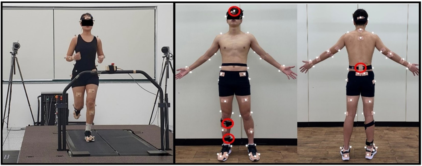
본 연구는 성별에 따른 신체의 운동학적 및 운동역학적 차이를 비교하기 위해 8대의 적외선 카메라(Oqus 300, Qualysis Track Manager, SWE; sampling rate: 100 Hz)와 지면반력기(force plate) 두 대가 내장된 트레드밀(Instrumented treadmill, Bertec, USA; sampling rate: 1,000 Hz), 3축 가속도계(accelero- meter; Width × Thickness: 1.9 cm × 1.1 cm, Ultium, Noraxon, USA; sampling rate: 1,000 Hz)를 활용하였다. 모든 연구 대상자들은 준비 운동 및 트레드밀 적응을 위해 3분간 트레드밀 위에서 달리기를 수행하였다(Figure 1 (A)). 각 연구 대상자들의 신발에 따른 충격 효과를 최소화하기 위해 N사의 운동화(Air Zoom Pegasus 39, Nike, USA)를 착용하였으며, 반사 마커 50개와 가속도계 네 채널을 부착하였다(Figure 1 (B), Lee, 2023; Lee, Ryu, Gil & Park, 2021; Ryu et al., 2021). 3축 가속도계는 신체의 정적인 해부학 자세를 기준으로 X 축은 위(+), 아래(-), Y 축은 오른쪽(+), 왼쪽(-), Z 축은 앞(-), 뒤(+)로 설정하였다. 모든 연구 대상자들은 트레드밀에서 2.8 m/s의 속도로 3분간 달리기를 수행하였으며, 이 중 마지막 1분만을 녹화하였다.

3. 자료 처리
본 연구의 위치와 지면반력 자료는 Qualisys Track Manager (Qualisys, SWE)를 통해 취득하였으며, 가속도계 자료는 MR 3.14 (Noraxon, USA)를 사용하여 취득하였다. 위치와 지면반력 자료는 Analog-digital board를 통해 동조하였고, 가속도계 자료는 MATLAB R2019a (MathWorks, USA)를 통해 동조하였다. 본 연구의 실험 절차에 따라 수집된 3차원 위치, 지면반력, 가속도계 자료는 취득 과정에서 발생하는 오차(random error)를 최소화하기 위해 2차 저역 통과 필터(Butterworth 2nd order low-pass filter)를 사용하였으며, 이때 차단 주파수(cut-off frequency)는 운동학적 자료 10 Hz, 운동역학적 및 가속도계 자료는 100 Hz로 설정하였다(Stergiou, Giakas, Byrne & Pomeroy, 2002). 분석 시점 및 구간은 주동발인 오른발의 착지 시점과 이지 순간, 충격 시점 그리고 디딤기로 나누어 분류하였으며, 1분 녹화 중 일정한 패턴을 보이는 20 stride를 선정하여 분석하였다. 하지 관절 및 몸통 분절의 각도, 가동 범위, 각속도와 관절 모멘트는 Visual3D (C-motion, USA)를 통해 산출하였으며, 보행 매개변수, 지면반력, 충격 가속도는 MATLAB R2019a를 통해 산출하였다.
4. 분석 변인
달리기 시 성별에 따른 생체역학적 변화를 비교하기 위해 보행 매개변수(gait parameter)와 3차원 하지 관절 및 몸통 분절 각도(lower extremity joint & trunk segment angle), 가동 범위(range of motion), 각속도(angular velocity), 지면반력(ground reaction force), 관절 모멘트(joint moment), 충격 가속도(impact acceleration)를 비교 · 분석하였다. 보행 매개변수는 디딤기 시간(contact time), 보장(step length), 보빈도(step frequency)를 산출하였다. 또한, 디딤기 시간을 전체 달리기 주기의 시간 대비 비율로, 보장을 피험자 개개인의 신장으로 표준화한 값도 추가로 산출하였다. 각도와 모멘트의 방향 설정은 <Table 1>과 같이 설정하였다. 지면반력은 수직 지면반력의 첫 번째 최댓값을 기준으로 최대 수직 충격력과 충격 부하율을 산출하였으며, Y 축 지면반력을 통해 제동력과 추진력을 산출하였다(Figure 2). 지면반력과 모멘트는 체중으로 나누어 표준화하였다. 충격 가속도는 경골에 부착된 센서의 수직, 수평 합성, 합성 가속도와 충격 흡수율을 산출하였으며, <Formula 1-2>
Formula 1. Resultant acceleration
Formula 2. Shock attenuation

|
|
X asis |
Y axis |
Z axis |
|
Ankle |
Dorsi-flexion
(+) |
Inversion
(+) |
Adduction
(+) |
|
Plantar
flexion (-) |
Eversion (-) |
Abduction (-) |
|
|
Knee |
Flexion (+) |
Adduction
(+) |
Internal
rotation (+) |
|
Hip |
Extension (-) |
Abduction (-) |
External
rotation (-) |
|
Trunk |
Extension
(+) |
Right
flexion (+) |
Left
rotation (+) |
|
Flexion (-) |
Left flexion
(-) |
Right
rotation (-) |
|
|
|
|||
5. 통계 분석
달리기 시 성별에 따른 운동학적 및 운동역학적 단일 변수의 차이를 검증하기 위해 SPSS 25.0 (IBM, USA) 프로그램을 사용하여 Levene의 등분산 검정과 Shapiro-Wilk 정규성 검정을 수행하였고, 독립 표본 t-검정(independent t-test)을 실시하였다. 또한, 연속적인 구간 전체(waveform)에 대한 분석을 위해 MATLAB R2019a (MathWorks, USA) 프로그램을 사용하여 오픈 소스(open source code; www.spm1d.org)로 제공된 1D-SPM (one-dimensional statistical parametric mapping) 기 법을 활용한 독립 표본 t-검정을 실시하였다. 이때 통계적 유의수준은 α=.05로 설정하였다.
1. 보행 매개변수(gait parameters)
달리기 시 성별에 따른 보행 매개변수를 비교한 결과는 <Table 2>와 같다. 디딤기 시간과 보장 그리고 보빈도에서 성별 간 통계적으로 유의한 차이가 나타났다(p<.05).
|
Variable |
Female |
Male |
t (p) |
|
|
Contact time |
(s) |
0.25±0.02 |
0.28±0.02 |
-3.491 (.002*) |
|
(%) |
37.03±2.99 |
38.04±4.45 |
-0.732 (.470) |
|
|
Step distance |
(m) |
0.95±0.06 |
1.04±0.05 |
-4.489 (.001*) |
|
(%) |
58.94±3.07 |
59.41±3.85 |
-0.370 (.714) |
|
|
Step frequency |
(Hz) |
1.55±0.08 |
1.42±0.07 |
4.784 (.001*) |
|
*: Indicates significant difference (p<.05) |
||||
2. 관절 및 분절 각도(joint & segment angle)
달리기 시 성별에 따른 관절 및 분절 각도를 비교한 결과는 <Table 3>과 같다. 무릎관절은 관상면 최대 각도에서 성별 간 통계적으로 유의한 차이가 나타났고(p<.05), 엉덩관절은 시상면 가동 범위에서 성별 간 통계적으로 유의한 차이가 나타났으며(p<.05), 몸통 분절은 시상면 및 수평면 최대 각도와 수평면 가동 범위에서 성별 간 통계적으로 유의한 차이가 나타났다(p<.05).
|
Variable |
Female |
Male |
t (p) |
|
|
Ankle max angle (deg) |
SP |
21.85±2.48 |
21.88±2.93 |
-0.032 (.975) |
|
FP |
8.21±2.62 |
9.36±2.31 |
-1.276 (.212) |
|
|
RoM of ankle (deg) |
SP |
36.26±3.12 |
35.83±4.34 |
0.316 (.754) |
|
FP |
10.55±3.37 |
10.38±3.65 |
0.134 (.894) |
|
|
Knee max angle (deg) |
SP |
35.83±3.23 |
34.84±3.78 |
0.775 (.445) |
|
FP |
-4.68±2.34 |
-0.44±2.58 |
-4.722 (.001*) |
|
|
RoM of knee (deg) |
SP |
29.84±2.90 |
29.10±2.30 |
0.767 (.450) |
|
FP |
4.17±2.30 |
3.50±1.35 |
0.981 (.335) |
|
|
Hip max angle (deg) |
SP |
27.19±6.66 |
25.38±5.05 |
0.838 (.155) |
|
FP |
9.62±3.07 |
8.06±2.75 |
1.463 (.692) |
|
|
RoM of hip (deg) |
SP |
38.18±2.34 |
34.18±2.98 |
4.092 (.001*) |
|
FP |
9.25±2.73 |
9.55±2.45 |
-0.318 (.753) |
|
|
Trunk max angle (deg) |
SP |
-7.10±4.41 |
-10.58±4.12 |
2.238 (.033*) |
|
TP |
-10.89±3.56 |
-7.53±1.71 |
-3.288 (.004*) |
|
|
RoM of trunk (deg) |
SP |
4.57±1.19 |
4.47±1.17 |
0.239 (.813) |
|
TP |
22.20±3.87 |
16.20±2.13 |
5.252 (.001*) |
|
|
*: Indicates significant difference (p<.05) max: Maximal, RoM: Range of motion SP (sagittal plane)-Ankle: Dorsi-flexion (+)/Plantar flexion (-), Knee/Hip: Flexion
(+)/Extension (-),
Trunk: Extension (+)/Flexion (-), FP (frontal plane)-Ankle: Inversion (+)/Eversion (-), Knee/Hip: Adduction
(+)/Abduction (-),
TP (transverse plane)-Trunk: Left rotation (+)/Right rotation (-) |
||||
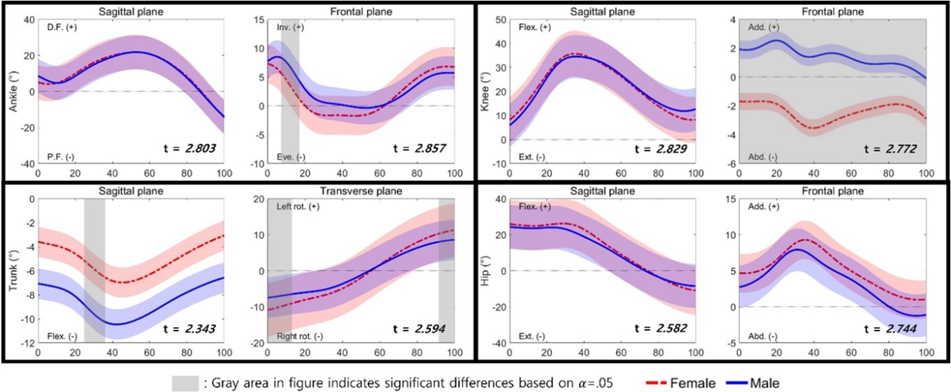
SPM 분석에 대한 결과는 <Figure 3>과 같다. 발목관절은 관상면 8~19%에서 성별 간 통계적으로 유의한 차이가 나타났고(p<.05), 무릎관절은 관상면 0~100%에서 성별 간 통계적으로 유의한 차이가 나타났으며(p<.05), 몸통 분절은 시상면 25~36%와 수평면 0~14% 및 93~100%에서 성별 간 통계적 으로 유의한 차이가 나타났다(p<.05).

3. 관절 및 분절 각속도(joint & segment angular velocity)
달리기 시 성별에 따른 관절 및 분절 각속도를 비교한 결과는 <Table 4>와 같다. 발목관절은 시상면에서 성별 간 통계적으로 유의한 차이가 나타났고(p<.05), 무릎관절은 시상면 및 관상면에서 성별 간 통계적으로 유의한 차이가 나타났다(p<.05). 엉덩관절은 시상면에서 성별 간 통계적으로 유의한 차이가 나타났으며(p<.05), 몸통 분절은 수평면에서 성별 간 통계적으로 유의한 차이가 나타났다(p<.05).
|
Variable |
Female |
Male |
t (p) |
|
|
Ankle max velocity (deg/sec) |
SP |
-470.53±38.49 |
-402.98±52.15 |
-4.036 (.001*) |
|
FP |
-193.05±54.42 |
-178.58±59.92 |
-0.692 (.494) |
|
|
Knee max velocity (deg/sec) |
SP |
-272.89±44.50 |
-231.03±40.29 |
-2.701 (.012*) |
|
FP |
-71.84±19.72 |
-54.46±17.92 |
-2.526 (.017*) |
|
|
Hip max velocity (deg/sec) |
SP |
-323.04±49.93 |
-233.24±28.13 |
-6.069 (.001*) |
|
FP |
-97.14±32.54 |
-87.13±19.86 |
-1.017 (.318) |
|
|
Trunk max velocity |
SP |
-56.92±18.38 |
-54.73±10.98 |
-0.396 (.695) |
|
TP |
24.09±17.22 |
-1.09±13.73 |
4.428 (.001*) |
|
|
*: Indicates significant difference (p<.05), max: Maximal SP (sagittal plane)-Ankle: Dorsi-flexion (+)/Plantar flexion (-), Knee/Hip: Flexion
(+)/Extension (-),
Trunk: Extension (+)/Flexion (-), FP (frontal plane)-Ankle: Inversion (+)/Eversion (-), Knee/Hip: Adduction
(+)/Abduction (-),
TP (transverse plane)-Trunk: Left rotation (+)/Right rotation (-) |
||||
4. 운동역학적 변인(kinetical variables)
달리기 시 성별에 따른 운동역학적 변인을 비교한 결과는 <Table 5>과 같다. 수직 충격 부하율과 최대 제동력에서 성별 간 통계적으로 유의한 차이가 나타났고(p<.05), 모멘트는 발목관절 시상면, 무릎관절 시상면 및 관상면, 엉덩관절 시상면에서 성별 간 통계적으로 유의한 차이가 나타났다(p<.05).
|
Variable |
Female |
Male |
t (p) |
|
|
VIPF (BW) |
1.46±0.24 |
1.49±0.20 |
-0.364 (.719) |
|
|
VILR (BW/s) |
66.46±19.50 |
44.41±10.42 |
3.865 (.001*) |
|
|
PBF (BW) |
0.22±0.02 |
0.20±0.02 |
2.161 (.039*) |
|
|
PPF (BW) |
-0.32±0.05 |
-0.31±0.04 |
-0.511 (.614) |
|
|
Ankle max
moment (Nm/kg) |
SP |
-2.02±0.25 |
-2.22±0.21 |
2.377 (.025*) |
|
FP |
0.50±0.22 |
0.44±0.17 |
0.817 (.421) |
|
|
Knee max
moment (Nm/kg) |
SP |
-2.07±0.23 |
-2.38±0.30 |
3.179 (.004*) |
|
FP |
0.62±0.34 |
0.29±0.20 |
3.227 (.003*) |
|
|
Hip max
moment (Nm/kg) |
SP |
0.83±0.20 |
0.63±0.24 |
2.497 (.019*) |
|
FP |
-1.68±0.39 |
-1.71±0.44 |
0.206 (.838) |
|
|
*: Indicates significant
difference (p<.05) VIPF: Vertical impact peak
force, VILR: Vertical impact loading rate, PBF: Peak braking force, PPF: Peak
propulsive force SP (sagittal plane)-Ankle: Dorsi-flexion (+)/Plantar flexion (-), Knee/Hip: Flexion (+)/Extension (-), FP (frontal plane)-Ankle: Inversion (+)/Eversion (-),
Knee/Hip: Adduction (+)/Abduction (-) |
||||
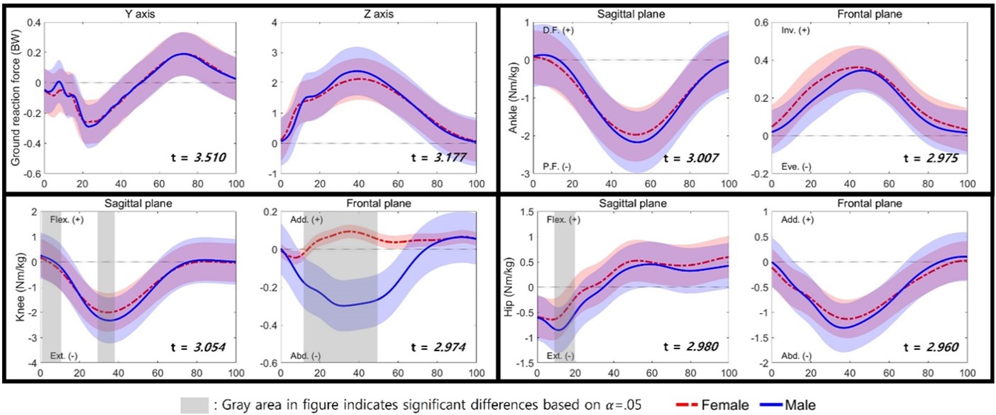
SPM 분석에 대한 결과는 <Figure 4>와 같다. 무릎관절은 시상면 0~11%와 31~39% 그리고 관상면 14~48%에서 성별 간 통계적으로 유의한 차이가 나타났으며(p<.05), 엉덩관절은 시상면 10~20%에서 성별 간 통계적으로 유의한 차이가 나타났다(p<.05).

5. 가속도 변인(acceleration variables)
달리기 시 성별에 따른 가속도 변인을 비교한 결과는 <Table 6>과 같다. 최대 수직 가속도는 모든 분절에서, 최대 수평 합성 가속도는 몸 쪽 정강뼈와 다섯 번째 허리뼈에서, 최대 합성 가속도는 모든 분절에서 성별 간 통계적으로 유의한 차이가 나타났다(Figure 5, p<.05). 먼 쪽 정강뼈에서 머리까지 충격 흡수율은 수평 합성 가속도에서 성별 간 통계적으로 유의한 차이가 나타났다(p<.05).
|
Variable |
Female |
Male |
t (p) |
|
|
Vertical Acc (g) |
D.T |
10.19±4.90 |
6.92±1.91 |
2.408 (.027*) |
|
P.T |
10.78±3.57 |
6.17±1.54 |
4.593 (.001*) |
|
|
5th
lumbar |
8.84±3.82 |
5.43±1.33 |
3.268 (.003*) |
|
|
Head |
3.06±0.61 |
2.54±0.33 |
2.882 (.007*) |
|
|
Horizontal resultant Acc (g) |
D.T |
7.62±3.83 |
5.87±2.42 |
1.499 (.145) |
|
P.T |
6.90±2.13 |
5.18±0.96 |
2.861 (.010*) |
|
|
5th
lumbar |
3.44±1.54 |
2.28±1.23 |
2.288 (.030*) |
|
|
Head |
0.85±0.19 |
1.03±0.34 |
-1.842 (.079) |
|
|
Resultant Acc (g) |
D.T |
11.81±5.49 |
8.36±2.53 |
2.207 (.039*) |
|
P.T |
12.59±3.98 |
7.54±1.47 |
4.613 (.001*) |
|
|
5th
lumbar |
12.58±4.91 |
5.72±1.82 |
5.072 (.001*) |
|
|
Head |
3.15±0.60 |
2.71±0.33 |
2.506 (.018*) |
|
|
D.T-Head SA (%) |
Vertical |
64.79±15.71 |
61.39±8.44 |
0.739 (.466) |
|
H.R |
86.16±7.31 |
79.50±9.68 |
2.126 (.042*) |
|
|
Resultant |
69.53±11.39 |
65.46±8.70 |
1.100 (.281) |
|
|
*: Indicates significant difference (p<.05) D.T: Distal tibia, P.T: Proximal tibia, H.R:
Horizontal resultant, SA: Shock attenuation |
||||

본 연구의 목적은 달리기 시 성별에 따른 운동학적 및 운동역학적 차이를 분석하고, 전신의 충격 특성을 파악하는 것이다.
1. 성별에 따른 보행 매개변수 차이
보행 매개변수의 차이를 비교한 결과, 보장은 여성이 남성에 비해 작게 나타난 반면, 보빈도는 여성이 남성에 비해 크게 나타났다. 이는 선행연구와 동일한 결과이며(Gehring et al., 2014), 본 연구에서 보장을 피험자 개개인의 신장으로 표준화한 데이터의 성별 간 차이가 나타나지 않은 것으로 미루어 보았을 때, 본 연구에 참여한 여성이 남성보다 평균적으로 약 14 cm 가량 신장이 작은 신체적 차이 때문이라고 판단된다.
2. 성별에 따른 하지 관절 및 몸통 분절 각도 차이
발목관절 각도의 차이를 비교한 결과, 3차원 운동축 모두 단일 변수에서 성별 간 차이가 나타나지 않았다. 이는 선행연구와 동일한 결과이지만(Sinclair et al., 2012), 본 연구에서 SPM 분석을 통해 연속 변수에서 남성의 안쪽 번짐(inversion) 각이 여성에 비해 크게 나타난 것을 추가로 확인할 수 있었다(관상면 8~19%; Figure 2). 선행연구에 따르면, 일반적으로 발목의 안쪽 번짐 최대 각은 디딤기의 15~30%에서 발생하며, 큰 안쪽 번짐 각이 발목 염좌와 같은 발목 부상의 잠재적 위험 요인이 될 수 있다고 하였다(Mousavi, Van Kouwenhove, Rajabi, Zwerver & Hijmans, 2021). 이처럼 연속 변수의 검증이 단일 변수의 검증에서 살펴볼 수 없었던 결과를 확인시켜 줄 수 있음을 시사하고 있으며, 상해와의 연관성을 발견하는 데 활용될 수 있을 것이라 생각된다.
무릎관절 각도의 차이를 비교한 결과, 선행연구와 마찬가지로(Sinclair et al., 2012) 최대 벌림(abduction) 각도가 남성에 비해 여성에서 크게 나타났다. 연속 변수의 검증에서도 단일 변수와 동일하게 여성의 벌림 각이 남성에 비해 크게 나타났다(관상면 0~100%; Figure 2). 선행연구에 따르면, 여성은 골반이 남성보다 상대적으로 넓어 무릎관절의 벌림을 초래하는 Q-angle 각이 크게 나타난다고 하였다(Kim, Jang & Choi, 2010). Han & Lim (2009)은 무릎관절의 벌림 움직임이 클수록 전방십자인대에 부하가 커진다고 보고하였다. 이는 무릎관절에서 수직, 수평 합성 및 합성 충격 가속도가 여성에 비해 남성에서 더 작게 나타난 본 연구의 결과와 일치한다고 볼 수 있으며, 이를 바탕으로 남성에 비해 여성이 달리기 시 무릎 상해에 노출될 확률이 높다고 예상된다.
몸통 분절 각도의 차이를 비교한 결과, 선행연구와 동일하게(Nagano, Sasaki, Higashihara & Ishii, 2016) 최대 굽힘 각은 여성에 비해 남성에서 크게 나타났고, 오른쪽 돌림 각과 수평면 가동 범위는 남성에 비해 여성에서 크게 나타났다. 연속 변수의 검증에서는 남성의 굽힘 각이 여성에 비해 크게 나타났고, 여성의 왼 · 오른쪽 돌림 각이 남성에 비해 크게 나타났다(시상면 25~36%; 수평면 0~14%, 93~100%; Figure 2). Apte et al. (2021)은 몸통 굽힘 각이 커질수록 발목관절 주변 근육에 과부하가 나타난다고 보고하였다. Teng & Powers (2015)의 연구에서는 몸통 굽힘 각이 커질수록 무릎의 폄 모멘트가 함께 증가하여 부상 위험이 높아질 수 있다고 하였다. 이는 해당 분절 움직임의 역할이 뚜렷함을 의미하며, 달리기 동작을 분석할 때 전신의 연결 체계를 확인할 필요가 있다고 판단된다. 또한, 남성의 발목 발바닥 굽힘 및 무릎 폄 모멘트가 여성에 비해 크게 나타났다는 본 연구의 결과와도 일치한다 볼 수 있다.
3. 성별에 따른 하지 관절 및 몸통 분절 각속도 차이
최대 각속도의 차이를 비교한 결과, 발목 발바닥 굽힘, 무릎 폄 및 벌림, 엉덩 폄, 몸통 왼쪽 돌림 각속도가 모두 여성이 남성에 비해 크게 나타났다. 선행연구에 따르면, 보빈도가 증가할수록 접촉 시간이 짧아지고, 다리의 움직임이 빨라지기 때문에 하지 관절의 각속도가 증가한다고 하였다(Clark, Meng & Stearne, 2020; Grimmer, Elshamanhory & Beckerle, 2020). 최근에는 무릎관절의 벌림에 관여하는 요인으로 체간 움직임이 주목을 받고 있다(Hewett, Torg & Boden, 2009; Kurihara et al., 2021; Sinclair et al., 2014). 앞서 논의한 내용과 같이 무릎관절의 벌림은 증가할수록 전방십자인대 손상의 위험성이 높아진다고 보고된 바 있다. 몸통과 하지는 해부학적으로 연결되어 있기 때문에 디딤기 동안 골반을 중심으로 서로 반대 방향으로 움직인다. 선행연구와 일맥상통하게 본 연구에서도 무릎관절 벌림과 체간 회전량이 함께 증가하는 경향을 확인할 수 있었다. 따라서 무릎관절의 벌림 움직임을 감소시키기 위해 이러한 상호작용을 고려해야 한다고 사료된다.
4. 성별에 따른 운동역학적 차이
지면반력 변인의 차이를 비교한 결과, 수직 충격 부하율과 최대 제동력이 남성에 비해 여성에서 크게 나타났다. 이는 Harrison, Ford, Myer & Hewett (2011)의 연구와 일치하는 결과이다. 선행연구들은 남성의 햄스트링 근육량이 여성보다 평균적으로 36% 더 많다고 보고하였다(Chumanov, Heiderscheit & Thelen, 2011; Janssen, Heymsfield, Wang & Ross, 2000; Malinzak et al., 2001). 따라서 남성에 비해 여성의 근육 활성화가 더 낮아 에너지를 덜 흡수하게 되고, 이로 인해 충격을 흡수하기 위해 부하율과 제동력이 증가한 것으로 생각된다.
무릎관절 최대 모멘트의 차이를 비교한 결과, 폄 모멘트가 여성에 비해 남성에서 크게 나타났다. 반면 모음 모멘트는 남성에 비해 여성에서 크게 나타났다. 연속 변수의 검증에서도 남성의 폄 모멘트가 여성에 비해 크게 나타났고, 여성의 모음 모멘트가 남성에 비해 크게 나타났다(시상면 0~11%, 31~39%; 관상면 14~48%; Figure 2). Gehring et al. (2014)은 남성에 비해 여성의 무릎 모음 모멘트가 크게 나타났으며, 그 원인으로 착지 시 무릎 벌림 각이 커져 관절이 내측으로 회전하려는 경향이 커지는 가능성을 제시하였다. 본 연구에서도 여성의 무릎 벌림 각과 충격 가속도가 크게 나타났으며, 이는 선행연구의 주장을 뒷받침한다. 따라서 남성에 비해 여성에서 지속적 관절 부하의 전달로 인해 퇴행성 골관절염이 유발될 확률이 상대적으로 높다고 생각된다.
5. 성별에 따른 부하와 충격의 차이
가속도 변인의 차이를 비교한 결과, 경골의 원위, 경골의 근위, 다섯 번째 허리뼈, 머리 분절 모두 최대 합성 가속도가 남성에 비해 여성에서 크게 나타났다. 신체 원위부에서 근위부로 올라갈수록 가속도의 크기가 감소된다는 선행연구의 결과와는 달리(Lee, 2023; Giandolini et al., 2016; Lucas-Cuevas, Encarnación-Martínez, Camacho-García, Llana-Belloch & Pérez-Soriano, 2017; Park et al., 2022; Ryu et al., 2021), 본 연구에서는 여성의 경우 경골의 원위보다 경골의 근위에서 가속도의 크기가 증가하였다. 이를 통해 남성은 발목에서, 여성은 무릎에서 더 큰 충격을 받는다고 생각되며, 남성의 발목 발바닥 굽힘 모멘트 및 여성의 무릎 모음 모멘트가 크게 나타나는 경향과 일치하는 것을 확인하였다. 따라서 관절 모멘트 대신 가속도계를 통해 관절의 부하를 간접적으로 측정할 수 있다고 사료된다. 먼 쪽 정강뼈부터 머리까지의 충격 흡수율은 수평 합성 가속도에서 여성이 남성에 비해 크게 나타났다. 이는 남성이 여성보다 발목관절에서 부하가 크다는 것을 의미하며, 각 분절에서의 충격 쇼크가 여성이 남성에 비해 크게 발생되었기 때문에 여성이 더 많은 충격 쇼크를 흡수한 것으로 판단된다.
본 연구를 통해 달리기 시 성별에 따른 생체역학적 차이를 확인하였다. 동일한 달리기 속도에서 남성의 경우 여성에 비해 신체 전체가 받는 충격력이 작게 나타나지만, 몸통을 앞으로 기울임에 따라 발목관절의 부하가 커져 발목 관련 부상을 입을 확률이 높아질 수 있다. 여성의 경우 해부학적 구조로 인해 무릎관절이 받는 충격력이 커져 무릎 관련 부상을 입을 확률이 높아질 수 있다. 따라서 이러한 손상을 방지하기 위해 발목 및 무릎 보호대를 착용하거나, 신발 안에 적절한 경도의 인솔을 넣어 충격을 흡수하는 것이 좋을 것으로 생각된다. 또한, 코어 근육을 강화시켜 체간 안정성을 높이는 것도 중요하다. 이를 통해 성별에 따라 달리 발생하는 달리기 부상 위험을 줄일 수 있을 것으로 사료된다.
References
1. Aghanezhad, H., Ahmadi, M., Noroozi, A. & Rahnama, N. (2022). Biomechanics of running: A special reference to the comparisons of wearing boots and running shoes. PLOS ONE, 17(7), e0270496.
Google Scholar
2. Apte, S., Prigent, G., Stöggl, T., Martínez, A., Snyder, C., Gremeaux-Bader, V. & Aminian, K. (2021). Biomechanical response of the lower extremity to running-induced acute fatigue: a systematic review. Frontiers in Physiology, 12, 646042.
3. Boyer, K. A., Freedman Silvernail, J. & Hamill, J. (2017). Age and sex influences on running mechanics and coordination variability. Journal of Sports Sciences, 35(22), 2225-2231.
Google Scholar
4. Cho, J. H. & Kim, R. B. (2015). The Effect of Restrict Movement of Knee Flextion during Landing on Impact Absorption Mechanism. Journal of Sport and Leisure Studies, 62, 815-824.
5. Chumanov, E. S., Heiderscheit, B. C. & Thelen, D. G. (2011). Hamstring musculotendon dynamics during stance and swing phases of high speed running. Medicine and Science in Sports and Exercise, 43(3), 525.
Google Scholar
6. Clark, K. P., Meng, C. R. & Stearne, D. J. (2020). 'Whip from the hip': thigh angular motion, ground contact mechanics, and running speed. Biology Open, 9(10), bio053546.
Google Scholar
7. Clermont, C. A., Benson, L. C., Osis, S. T., Kobsar, D. & Ferber, R. (2019). Running patterns for male and female competitive and recreational runners based on accelerometer data. Journal of Sports Sciences, 37(2), 204-211.
Google Scholar
8. Dozza, M., Horak, F. & Chiari, L. (2004). Audio Biofeedback of Trunk Accelerations improves balance in subjects with bilateral vestibular loss. In Proc. 28th Annual Meeting of the American Society of Biomechanics.
Google Scholar
9. Edwards, W. B., Derrick, T. R. & Hamill, J. (2012). Musculo- skeletal attenuation of impact shock in response to knee angle manipulation. Journal of Applied Biomechanics, 28(5), 502-510.
Google Scholar
10. Fleming, P., Young, C., Dixon, S. & Carré, M. (2010). Athlete and coach perceptions of technology needs for evaluating running performance. Sports Engineering, 13(1), 1-18.
Google Scholar
11. Gehring, D., Mornieux, G., Fleischmann, J. & Gollhofer, A. (2014). Knee and hip joint biomechanics are gender-specific in runners with high running mileage. International Journal of Sports Medicine, 35(02), 153-158.
Google Scholar
12. Giandolini, M., Horvais, N., Rossi, J., Millet, G. Y., Samozino, P. & Morin, J. B. (2016). Foot strike pattern differently affects the axial and transverse components of shock acceler- ation and attenuation in downhill trail running. Journal of Biomechanics, 49(9), 1765-1771.
Google Scholar
13. Grimmer, M., Elshamanhory, A. A. & Beckerle, P. (2020). Human lower limb joint biomechanics in daily life activities: a literature based requirement analysis for anthropomorphic robot design. Frontiers in Robotics and AI, 7, 13.
Google Scholar
14. Gruber, A. H., Boyer, K. A., Derrick, T. R. & Hamill, J. (2014). Impact shock frequency components and attenuation in rearfoot and forefoot running. Journal of Sport and Health Science, 3(2), 113-121.
Google Scholar
15. Han, K. H. & Lim, B. O. (2009). Mechanism and risk factors of anterior cruciate ligament injuries in female athletes. The Official Journal of the Korean Academy of Kinesiology, 11(3), 61-83.
Google Scholar
16. Han, S. H. & Kwon, J. H. (2015). Gender differences in Dynamic Q-angle during walking with Motion Capture. Proceedings of the Society of CAD/CAM Conference, 812-817.
17. Harrison, A. D., Ford, K. R., Myer, G. D. & Hewett, T. E. (2011). Sex differences in force attenuation: a clinical assessment of single-leg hop performance on a portable force plate. British Journal of Sports Medicine, 45(3), 198-202.
Google Scholar
18. Hennig, E. M. (2001). Gender differences for running in athletic footwear. In Fifth Symposium on Footwear Biomechanics. ETH Zürich, Switzerland.
Google Scholar
19. Hewett, T. E., Torg, J. S. & Boden, B. P. (2009). Video analysis of trunk and knee motion during non-contact ACL injury in female athletes: lateral trunk and knee abduction motion are combined components of the injury mechanism. British Journal of Sports Medicine, 1-26.
Google Scholar
20. Hooker, S. P., Feeney, A., Hutto, B., Pfeiffer, K. A., McIver, K., Heil, D. P. & Blair, S. N. (2011). Validation of the actical activity monitor in middle-aged and older adults. Journal of Physical Activity and Health, 8(3), 372-381.
Google Scholar
21. Hreljac, A. (2004). Impact and overuse injuries in runners. Medicine & Science in Sports & Exercise, 36(5), 845-849.
Google Scholar
22. Janssen, I., Heymsfield, S. B., Wang, Z. & Ross, R. (2000). Skeletal muscle mass and distribution in 468 men and women aged 18~88 yr. Journal of Applied Physiology, 89, 81-88.
Google Scholar
23. Kang, H. B., Kim, G., Kim, H., Han, S. R., Chae, D. J., Song, H. J. & Kim, D. W. (2013). Cerebrolysin attenuates astrocyte activation following repetitive mild traumatic brain injury: implications for chronic traumatic encephalopathy. Journal of Life Science, 23(9), 1096-1103.
Google Scholar
24. Kim, H. K., Mirjalili, S. A., Zhang, Y., Xiang, L., Gu, Y. & Fernandez, J. (2024). Effect of gender and running experience on lower limb biomechanics following 5 km barefoot running. Sports Biomechanics, 23(1), 95-108.
Google Scholar
25. Kim, J. N. (2020). Correlation Analysis between Joint Stiffness and Biomechanical Load during Running. The Korean Journal of Sport, 18(3), 1369-1377.
26. Kim, T. S., Jang, J. W. & Choi, J. H. (2010). Effect of Rehabilitation Exercise during 8 Weeks on Lower Extremity Muscular-function and Function Score in Women's Soccer Player with Patellofemoral Pain Syndrome. Journal of Sport and Leisure Studies, 42(2), 1117-1126.
27. Kugler, F. & Janshen, L. (2010). Body position determines propulsive forces in accelerated running. Journal of Bio- mechanics, 43(2), 343-348.
Google Scholar
28. Kumahara, H., Ishii, K. & Tanaka, H. (2006). Physical activity monitoring for health management: practical techniques and methodological issues. International Journal of Sport and Health Science, 4(2), 380-393.
Google Scholar
29. Kurihara, Y., Ohsugi, H., Karasuno, H., Tagami, M., Matsuda, T. & Fujikawa, D. (2021). Trunk rotation enhances movement of the knee abduction angle while running among female collegiate middle-and long-distance runners. Journal of Human Sport and Exercise, 17(4), 750-760.
Google Scholar
30. Lee, Y. S. (2023). Effects of Heel Strike Pattern on Full-body Impact during Running. Un- published Doctor's Disser- tation. Graduate School of Korea National Sport University.
31. Lee, Y. S., Ryu, S., Gil, H. J. & Park, S. K. (2021). Impact and Shock Attenuation of the Runners with and without Low Back Pain. Korean Journal of Sport Biomechanics, 31(1), 16-23.
Google Scholar
32. Lucas-Cuevas, A. G., Encarnación-Martínez, A., Camacho-García, A., Llana-Belloch, S. & Pérez-Soriano, P. (2017). The loca- tion of the tibial accelerometer does influence impact acceleration parameters during running. Journal of Sports Sciences, 35(17), 1734-1738.
Google Scholar
33. Malinzak, R. A., Colby, S. M., Kirkendall, D. T., Yu, B. & Garrett, W. E. (2001). A comparison of knee joint motion patterns between men and women in selected athletic tasks. Clinical Biomechanics, 16(5), 438-445.
Google Scholar
34. Matthews, C. E., Jurj, A. L., Shu, X. O., Li, H. L., Yang, G., Li, Q. & Zheng, W. (2007). Influence of exercise, walking, cycling, and overall nonexercise physical activity on mortality in Chinese Female. American Journal of Epidemiology, 165(12), 1343-1350.
Google Scholar
35. Mayagoitia, R. E., Nene, A. V. & Veltink, P. H. (2002). Accelero- meter and rate gyroscope measurement of kinematics: an inexpensive alternative to optical motion analysis systems. Journal of Biomechanics, 35(4), 537-542.
Google Scholar
36. Mazzà, C., Iosa, M., Picerno, P. & Cappozzo, A. (2009). Gender differences in the control of the upper body accelerations during level walking. Gait & Posture, 29(2), 300-303.
Google Scholar
37. Meardon, S. A. & Derrick, T. R. (2014). Effect of step width manipulation on tibial stress during running. Journal of Biomechanics, 47(11), 2738-2744.
Google Scholar
38. Mousavi, S. H., Van Kouwenhove, L., Rajabi, R., Zwerver, J. & Hijmans, J. M. (2021). The effect of changing foot pro- gression angle using real-time visual feedback on rearfoot eversion during running. PLoS One, 16(2), e0246425.
Google Scholar
39. Nagano, Y., Sasaki, S., Higashihara, A. & Ishii, H. (2016). Gender differences in trunk acceleration and related posture during shuttle run cutting. International Biomechanics, 3(1), 33-39.
Google Scholar
40. Oh, Y. J. & Lee, C. M. (2009). The Study on 3-Axes Acceleration Impact of Lower Limbs Joint during Gait. Journal of the Ergonomics Society of Korea, 28(3), 33-39.
Google Scholar
41. Park, S. H., Yoon, S. H., Park, S. K. & Ryu, J. S. (2018). Initial Contact Angle of the Foot Segment and GRF Com- ponents by the Gender Differences. The Korean Journal of Physical Education, 57(2), 625-633.
42. Park, S. K., Stefanyshyn, D., Ryu, S., Gil, H., Lee, Y. S., Kim, J. & Ryu, J. (2022). Comparisons of Age-Related Changes in Impact Characteristics Between Healthy Older and Younger Runners. International Journal of Precision Engineering and Manufacturing, 23(12), 1465-1476.
Google Scholar
43. Phinyomark, A., Hettinga, B. A., Osis, S. T. & Ferber, R. (2014). Gender and age-related differences in bilateral lower ex- tremity mechanics during treadmill running. PloS One, 9(8), e105246.
Google Scholar
44. Ryu, S., Lee, Y. S. & Park, S. K. (2021). Impact signal differences dependent on the position of accelerometer attachment and the correlation with the ground reaction force during running. International Journal of Precision Engineering and Manufacturing, 22, 1791-1798.
Google Scholar
45. Scheerder, J., Breedveld, K. & Danchev, A. (2014). Running Across Europe: The Rise and Size of One of the Largest Sport Markets. London: Palgrave Macmillan.
Google Scholar
46. Sinclair, J. K., Chockalingam, N. & Vincent, H. (2014). Gender differences in multi-segment foot kinematics and plantar fascia strain during running. Foot & Ankle, 7(4).
Google Scholar
47. Sinclair, J., Greenhalgh, A., Edmundson, C. J., Brooks, D. & Hobbs, S. J. (2012). Gender differences in the kinetics and kinematics of distance running: implications for foot- wear design. International Journal of Sports Science and Engineering, 6(2), 118-128.
Google Scholar
48. Stergiou, N., Giakas, G., Byrne, J. E. & Pomeroy, V. (2002). Fre- quency domain characteristics of ground reaction forces during walking of young and elderly females. Clinical Biomechanics, 17(8), 615-617.
Google Scholar
49. Takabayashi, T., Edama, M., Nakamura, M., Nakamura, E., Inai, T. & Kubo, M. (2017). Gender differences associated with rearfoot, midfoot, and forefoot kinematics during running. European Journal of Sport Science, 17(10), 1289-1296.
Google Scholar
50. Taunton, J. E., Ryan, M. B., Clement, D. B., McKenzie, D. C., Lloyd-Smith, D. R. & Zumbo, B. D. (2002). A retrospective case-control analysis of 2002 running injuries. British Journal of Sports Medicine, 36(2), 95-101.
Google Scholar
51. Teng, H. L. & Powers, C. M. (2015). Influence of trunk posture on lower extremity energetics during running. Medicine & Science in Sports & Exercise, 47(3), 625-630.
Google Scholar
52. Toth, A. P. & Cordasco, F. A. (2001). Anterior cruciate ligament injuries in the female athlete. Journal of Gender-Specific Medicine, 4(4), 25-34.
Google Scholar
53. Van Gent, R. N., Siem, D., Van Middelkoop, M., Van Os, A. G., Bierma-Zeinstra, S. M. A. & Koes, B. W. (2007). Incidence and determinants of lower extremity running injuries in long distance runners: a systematic review. British Journal of Sports Medicine, 41(8), 469-480.
Google Scholar
54. Winter, D. A. (2009). Biomechanics and Motor Control of Human Movement. Hoboken: John Wiley & Sons lnc.
Google Scholar
55. Harsted, S., Holsgaard-Larsen, A., Hestbaek, L., Boyle, E. & Lauridsen, H. H. (2019). Concurrent validity of lower ex- tremity kinematics and jump characteristics captured in pre-school children by a markerless 3D motion capture system. Chiropractic & Manual Therapies, 27(1), 1-16.
Google Scholar