Open Access, Peer-reviewed
eISSN 2093-9752
Open Access, Peer-reviewed
eISSN 2093-9752
Si-Hyun Yoo
Jong-Bin Kim
Ji-Seon Ryu
Suk-Hoon Yoon
Sang-Kyoon Park
http://dx.doi.org/10.5103/KJSB.2016.26.2.221 Epub 2016 July 17
Abstract
Objective: The purpose of this study was to investigate differences in gait parameters and symmetry between walking speed by using the Froude number and preferred walking speed.
Method: Fifty adults (age: 21.0 ± 1.7 years, body weight: 71.0 ± 9.2 kg, height: 1.75 ± 0.07 m, leg length: 0.89 ± 0.05 m) participated in this study. Leg length-applied walking speed was calculated by using the Froude number, defined as Fr = v2/gL, where v is the velocity, g is the gravitational acceleration, and L is the leg length. Video data were collected by using eight infrared cameras (Oqus 300, Qualysis, Sweden) and the Qualisys Track Manager software (Qualisys, Sweden), with a 200-Hz sampling frequency during two-speed walking (preferred walking speed [PS] and leg length-applied walking speed [LS]) on a treadmill (Instrumented Treadmill, Bertec, USA). The step length, stride length, support percentage, cadence, lower joint angle, range of motion (ROM), and symmetry index were then calculated by using the Matlab R2009a software.
Results: Step and stride lengths were greater in LS than in PS (p < 0.05). The right single-support percentage was greater in LS than in PS (p < 0.05). The hip joint angle at heel contact and toe-off were greater in LS than in PS (p < 0.05). The hip and knee joint ROM were greater in LS than in PS (p < 0.05).
Conclusion: Based on our findings, we suggest that increased walking speed had a significant effect on step length, stride length, support percentage, and lower joint ROM.
Keywords
Gait Symmetry Walking speed Froude number
Walking is the most basic mode of transportation of humans. It is a complex motion produced by the coordination and control of the upper and lower limb joints. Gait varies depending on the physical traits, habits, walking speed, and personality (Shin, Lee, & Kwon, 2008; Tirosh & Sparrow, 2005; Whittle, 1990).
So far, the symmetrical movements of both legs have been regarded as normal gait. However, even non-handicapped individuals can de- velop asymmetrical gait because of physical traits or differences in function between both feet (Sadeghi, Allard, Prince, & Labelle, 2000). Echeverria, Rodriguez, Velascol, and Alvarez-Ramirez (2010) reported that around 10% of cases of asymmetrical gait could be observed in non-handicapped individuals. Improper gait and habits can cause diseases in body structure such as bones and muscles (Moon, 2005; Scott & Winter, 1990). In particular, asymmetrical gait can cause body fatigue and can directly cause disabilities or diseases because abnormal loads during gait can travel all the way to the brain (Nigg, De Boer, & Fisher, 1995). Asymmetry produced while walking is used to evaluate gait. In their study, Perttunen, Anttila, Sodergard, Merikanto, and Komi (2004) examined the asymmetrical gait of patients with lower limb length discrepancies and reported that weight bearing was higher on the longer leg. In another study, Lugade, We, Jewett, Collis, and Chou (2010) reported that surgery improved asymmetrical gait in patients with hip osteoarthritis. Hyun and Ryew (2014) investigated the correlation be- tween heel height and gait in young female adults and reported that heel height was directly proportional to asymmetry and observed bi- lateral asymmetry. Roth, Mervitz, Mroczek, Dugan, and Suh in 1997; Patterson et al. in 2008; and Nam, Kim, and An in 2010 reported that bilateral asymmetry increases with decreased walking speed in stroke patients. Those authors proposed that asymmetry index should be con- sidered an assessment parameter of gait. The asymmetry magnitude in walking is important in the evaluation of gait and is used as an important criterion for preventing lesion and promoting rehabilitation. Accordingly, efforts to reduce gait asymmetry are required.
Speed directly induces asymmetry and changes in gait (Murray, 1967; Vaughan, Toit, & Roffey, 1987; Winter & White, 1987). A previous study by Murray in 1967 analyzed differences between regular and fast walking, and investigated the effects of walking speed on lower limb joints. In their study in 1978, Crowinshield, Brand, and Johnston reported that stride length, leg strength, and hip joint rotational force and range of motion increase as walking speed increases. In addition, Kim, Lee, and Jin (2000) and Vaughan, Langerak, and O'Malley (2003) reported that increased walking speed increases lower limb range of motion and directly affects lower limb movements. Therefore, walking speed and gait asymmetry, which directly cause changes in gait pattern and body movements, should be further investigated.
Walking speed is most commonly measured by using the preferred walking speed method and walking speed measurements that reflect lower limb lengths. The preferred walking speed method measures speed by dividing the distance walked in a straight line on a flat surface by the time it took to walk the distance (Choi, Kang, & Tack, 2011; Soong, Lovie-Kitchin, & Brown, 2000). By contrast, walking speed meas- urements that reflect lower limb lengths involve the use of the Froude number, which is derived from the pendulum model, and measure walking speed that reflects both lower limb lengths and gravitational acceleration (Alexander & Jayes, 1983; Choi et al., 2011; England & Granata, 2007; Vaughan & O'Malley, 2005). The fundamental research by Choi et al. in 2011 investigated walking speed by using the two aforementioned methods and reported significant differences between the preferred walking speed method and the walking speed measure- ments that reflect lower limb lengths. The differences were inferred to be due to the fact that preferred walking speed largely varies depending on individual physical traits, habits, and personality. Hof (1996) and Lee (2014) reported that it is difficult to determine whether differences in gait pattern, including walking speed, are caused by sex-related factors or physical traits such as weight and height. The studies also men- tioned that physical traits such as weight and height should always be converted to standardized values prior to comparison. The present study is significant because it investigated differences between the most commonly used preferred walking speed method and walking speed measurements that reflect not only lower limb lengths but also gait parameters and asymmetry magnitude. The purpose of this study was to investigate the asymmetry and differences in gait parameters between the preferred walking speed and leg length-applied walking speed.
1. Subjects
Taking into account effect size (0.5), statistical power (0.95), and p value (0.05), 50 adult men in their twenties who had no history of orthopedic diseases were selected as subjects. The anthropometric information of the participants is shown in (Table 1).
|
n |
Age |
Body
weight |
Height |
Leg
length |
|
50 |
21.0 ± 1.7 |
71.0 ± 9.2 |
1.75 ± 0.07 |
0.89 ± 0.05 |
2. Experimental procedure
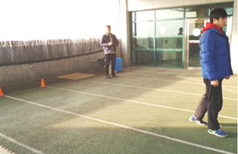
To investigate differences between preferred walking speed and leg length-applied walking speed, both walking speeds were calculated. As shown in (Figure 1), preferred walking speed was determined by having the participants walk a 50-m-long straight path 3 times in their usual walking speed and by calculating their average speeds (Choi et al., 2011; Soong et al., 2000). Leg length-applied walking speed was deter- mined by using gravitational acceleration, leg lengths, and a Froude number of 0.25. Leg length was measured by measuring the height of the greater trochanter in the standing position (Alexander & Jayes, 1983; Choi et al., 2011; England & Granata, 2007; Vaughan & O'Malley, 2005). The Froude number was set at 0.25, which was the optimal Froude number for walking speed calculation according to Vaughan and O'Malley (2005).
Fr = Froude number, v = walking speed,
g = gravitational acceleration, and L = leg length.
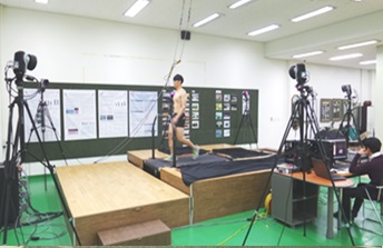
The participants walked on a treadmill (Instrumented Treadmill, Bertec, USA) at two calculated walking speeds (Figure 2). Reflective markers were placed on the joints (left acromion, right acromion, left greater trochanter, right greater trochanter, left femoral condyles, right femoral condyles, left malleolus, right malleolus, left heel bone, right heel bone, left first phalanges, and right first phalanges). Eight infrared cameras (Oqus 300, Qualisys, Sweden) were used to digitally record the move- ment of the reflective markers while the participants walked. Initially, the participants were given 5 minutes to adapt to the treadmill, and the strides on the succeeding 30 minutes were recorded. The recording speed was set to 200 Hz.
3. Data processing
The Qualisys Track Manager software (Qualisys, Sweden) and Matlab R2014b software (The MathWorks, Inc., USA) were used to analyze gait parameters and asymmetry. The nonlinear transformation (NLT) method was used to convert the reflective marker coordinates into three-dimensional coordinates. To eliminate noise, the recordings were smoothed by using a Butterworth second-order low-pass filter at a cutoff frequency of 6 Hz.
4. Analysis variables
1) Gait parameters
The step length, stride length, stride time, double- and single-sup- port percentage, two-dimensional angles, and range of motion of the lower limb joints and cadence, which were produced while walking on the treadmill at the two walking speeds, were calculated. The direction of the lower limb joint angle was classified as either counterclockwise (+) and clockwise (-).
2) Symmetry index
Excluding cadence, all the variables in both feet were used to calcu- late the asymmetry index (Kim & Eng, 2003; Robinson, Herzog, & Nigg, 1987). A symmetry index (SI) close to 0% signifies bilateral symmetry, with a range of up to 200% (Kim & Eng, 2003).
5. Statistical processing
In this study, SPSS Ver. 18.0 software (IBM, USA) and paired t test were used to investigate the differences between preferred walking speed and leg length-applied walking speed. The significance level was set to 0.05.
Gait variables and differences in lower limb joint angles between the preferred walking speed and leg length-applied walking speed were analyzed. To determine the magnitude of asymmetry, walking speed, step length, stride length, stride time, double- and single-support per- centage, and leg joint range of motion were analyzed and the asym- metry index was calculated.
1. Gait parameters
Although the preferred walking speed was 1.43 ± 0.16 m/s and the leg length-applied walking speed was 1.48 ± 0.04 m/s, no statistically significant differences were observed (Table 2). The step length of the right foot was 0.62 ± 0.05 m at the preferred walking speed and 0.64 ± 0.04 m at the leg length-applied walking speed. The step length of the left foot was 0.62 ± 0.05 m and 0.64 ± 0.04 m at the preferred and leg length-applied walking speeds, respectively. Significant differ- ences were observed between the right (p = 0.007) and left feet (p = 0.000). The stride lengths of both the right and left feet were 1.24 ± 0.10 m at the preferred walking speed and 1.28 ± 0.08 m at the leg length-applied walking speed, indicating significant differences between the right (p = 0.003) and left feet (p = 0.002). However, no significant differences in the number of strides were observed between the left and right feet. The single-support percentages of the right foot were 32.39% ± 2.73% and 33.65% ± 3.51% for the preferred and leg length-applied walking speeds, respectively, which are significantly different at p = 0.048.
2. Lower limb joint angle
When the heel contacted the ground, the joint angle of the right hip joint was 25.52° ± 3.13° at the preferred walking speed and 26.17° ± 2.76° at the leg length-applied walking speed (Table 3). The joint angle of the left hip joint was 25.21° ± 3.40° at the preferred walking speed and 26.77° ± 3.61° at the leg length-applied walking speed, with sig- nificant differences between right (p = 0.001) and left hip joints (p = 0.044). The joint angle of the left knee joint was -5.99° ± 4.81° at the Table 2. Descriptive information for gait variables Gait variables Preferred walking speed Leg length-applied walking speed (Froude number) t P Walking speed (m/s) 1.43 ± 0.16 1.48 ± 0.04 -1.846 0.071 Right step length (m) 0.62 ± 0.05 0.64 ± 0.04 -2.810 0.007* Left step length (m) 0.62 ± 0.05 0.64 ± 0.04 -3.776 0.000* Right stride length (m) 1.24 ± 0.10 1.28 ± 0.08 -3.133 0.003* Left stride length (m) 1.24 ± 0.10 1.28 ± 0.08 -3.291 0.002* Right stride time (sec) 1.13 ± 0.21 1.10 ± 0.12 0.833 0.409 Left stride time (sec) 1.13 ± 0.21 1.10 ± 0.11 0.984 0.330 Right double support (%) 16.52 ± 3.21 15.95 ± 1.18 1.298 0.201 Right single support (%) 32.39 ± 2.73 33.65 ± 3.51 -2.030 0.048* Left double support (%) 14.36 ± 3.60 13.39 ± 1.45 1.785 0.080 Left single support (%) 36.73 ± 2.26 37.01 ± 3.02 -0.560 0.578 Cadence (step/min) 109.82 ± 10.24 111.57 ± 6.29 -1.243 0.220 *Statistically significant difference between the preferred and leg length-applied walking speeds (Froude number) at a level of 0.05. Bolded Number indicates significant differences between two conditions. preferred walking speed and -5.50° ± 5.00° at the leg length-applied walking speed, indicating significant differences (p = 0.026). As the heel left from the ground, the joint angle of the right hip joint was 1.53° ± 4.01° at the preferred walking speed and 0.82° ± 4.06° at the leg length-applied walking speed. A significant difference was observed only in the right hip joint angle as the heel left from the ground (p = 0.032). The right leg joint range of motion at the standing phase was 36.51° ± 3.39° at the preferred walking speed and 37.89° ± 2.88° at the leg length-applied walking speed. The left leg joint range of motion at the standing phase was 36.48° ± 3.68° at the preferred walking speed and 37.54° ± 3.08° at the leg length-applied walking speed. Significant differences were observed for the right (p = 0.000) and left leg joints (p = 0.011). The right leg joint range of motion at the swing phase was 28.75° ± 3.86° at the preferred walking speed and 30.21° ± 4.04° at the leg length-applied walking speed. The left leg joint range of motion at the swing phase was 27.80° ± 3.97° at the preferred walking speed and 29.28° ± 3.82° at the leg length applied walking speed. Significant differences were observed for the right (p = 0.003) and left leg joints (p = 0.005). The right knee joint range of motion was 65.69° ± 3.78° at the preferred walking speed and 67.86° ± 3.75° at the leg length-applied walking speed. The left knee joint range of motion was 65.59° ± 4.06° at the preferred walking speed and 67.57° ± 3.80° at the leg length-applied walking speed. Significant differences were observed for both the right and left leg joints (p = 0.000).
|
Gait variables |
Preferred walking speed |
Leg length-applied walking speed (Froude number) |
t |
P |
|
Walking speed (m/s) |
1.43 ± 0.16 |
1.48 ± 0.04 |
-1.846 |
0.071 |
|
Right step length (m) |
0.62 ± 0.05 |
0.64 ± 0.04 |
-2.810 |
0.007* |
|
Left step length (m) |
0.62 ± 0.05 |
0.64 ± 0.04 |
-3.776 |
0.000* |
|
Right stride length (m) |
1.24 ± 0.10 |
1.28 ± 0.08 |
-3.133 |
0.003* |
|
Left stride length (m) |
1.24 ± 0.10 |
1.28 ± 0.08 |
-3.291 |
0.002* |
|
Right stride time (sec) |
1.13 ± 0.21 |
1.10 ± 0.12 |
0.833 |
0.409 |
|
Left stride time (sec) |
1.13 ± 0.21 |
1.10 ± 0.11 |
0.984 |
0.330 |
|
Right double support (%) |
16.52 ± 3.21 |
15.95 ± 1.18 |
1.298 |
0.201 |
|
Right single support (%) |
32.39 ± 2.73 |
33.65 ± 3.51 |
-2.030 |
0.048* |
|
Left double support (%) |
14.36 ± 3.60 |
13.39 ± 1.45 |
1.785 |
0.080 |
|
Left single support (%) |
36.73 ± 2.26 |
37.01 ± 3.02 |
-0.560 |
0.578 |
|
Cadence (step/min) |
109.82 ± 10.24 |
111.57 ± 6.29 |
-1.243 |
0.220 |
3. Bilateral SI
The magnitude of asymmetry was calculated by using step length, stride length, stride time, double- and single-support percentages, and ranges of motion of the hip, knee, and the ankle joints at the standing and swing phases. No statistically significant differences in the SIs of all the variables were observed between the preferred and leg length-applied walking speeds (Table 4).
The purpose of this study was to investigate differences in lower limb joint angles, bilateral asymmetry, and gait parameters between preferred and leg length-applied walking speed.
The preferred walking speed was 1.43 ± 0.16 m/s, and the leg length-applied walking speed was 1.48 ± 0.04 m/s. Although no statistically significant differences were observed, leg length-applied walking speed was relatively greater than the preferred walking speed. The preferred walking speed determined in this study was similar to the 1.39 m/s reported by Skinner and Barrack in 1990 and the 1.43 m/s reported by Perry in 1992. In their study, Choi, Kang, Mun, Bang, and Tack (2010) analyzed kinematic differences in gait between adults and seniors and reported preferred a walking speed of 1.44 m/s. As the average height of subjects in the above-mentioned study was 174.3 cm, the preferred walking speed of 1.43 m/s observed in the present study is inferred to be within the standard speed range. However, the study by Kim and Yoon in 2009, which aimed to collect standardized data on Koreans' normal walking, reported that the average walking speed of 73 adult participants was 1.21 m/s, which is relatively slow. This result could be due to the fact that their average height was 166.7 cm and the average leg length was 85.4 cm. Therefore, we can infer that height and leg length directly affect walking speed. Step and stride lengths were observed to be greater at the leg length-applied walking speed than at the preferred walking speed, which coincides with the results of the study by Crowinshield et al. in 1978. The difference in step and stride lengths could be attributed to the differences in walking speed. At the leg length-applied speed, double-support percentage was observed to be relatively lower, whereas single-support percentage was higher. We can infer that the relatively faster leg length walking speed causes an increase in step and stride lengths due to a longer support time.
|
Lower joint angle |
Preferred walking speed |
Leg length-applied walking
speed (Froude number) |
t |
p |
|
Angle at heel contact |
|
|
|
|
|
Right hip |
25.52 ± 3.13 |
26.17 ± 2.76 |
-3.387 |
0.001* |
|
Right knee |
-5.99 ± 4.81 |
-5.50 ± 5.00 |
-2.289 |
0.026* |
|
Right ankle |
-5.94 ± 4.14 |
-5.70 ± 3.86 |
-0.582 |
0.563 |
|
Left hip |
25.21 ± 3.40 |
26.77 ± 3.61 |
-2.072 |
0.044* |
|
Left knee |
-5.10 ± 4.81 |
-5.92 ± 4.91 |
1.099 |
0.277 |
|
Left ankle |
-5.90 ± 4.09 |
-5.91 ± 3.88 |
0.065 |
0.949 |
|
Angle at toe-off |
|
|
|
|
|
Right hip |
1.53 ± 4.01 |
0.82 ± 4.06 |
2.214 |
0.032* |
|
Right knee |
-52.34 ± 4.91 |
-52.34 ± 5.34 |
-0.005 |
0.996 |
|
Right ankle |
-24.67 ± 4.50 |
-24.04 ± 4.35 |
-1.374 |
0.176 |
|
Left hip |
1.99 ± 4.77 |
2.18 ± 4.62 |
-0.233 |
0.817 |
|
Left knee |
-53.33 ± 5.29 |
-54.31 ± 5.34 |
1.184 |
0.242 |
|
Left ankle |
-25.21 ± 5.02 |
-24.77 ± 4.54 |
-1.005 |
0.320 |
|
Ranges of motion (ROM) at |
|
|
|
|
|
Right hip |
36.51 ± 3.39 |
37.89 ± 2.88 |
-3.778 |
0.000* |
|
Right knee |
46.76 ± 4.25 |
47.26 ± 4.87 |
-1.012 |
0.317 |
|
Right ankle |
28.54 ± 3.41 |
28.76 ± 3.59 |
-0.603 |
0.549 |
|
Left hip |
36.48 ± 3.68 |
37.54 ± 3.08 |
-2.631 |
0.011* |
|
Left knee |
48.78 ± 4.54 |
48.57 ± 4.56 |
0.428 |
0.671 |
|
Left ankle |
29.40 ± 3.70 |
29.94 ± 3.85 |
-1.860 |
0.069 |
|
ROM at swing phase |
|
|
|
|
|
Right hip |
28.75 ± 3.86 |
30.21 ± 4.04 |
-3.166 |
0.003* |
|
Right knee |
65.69 ± 3.78 |
67.86 ± 3.75 |
-7.147 |
0.000* |
|
Right ankle |
25.48 ± 4.83 |
25.97 ± 3.92 |
-0.864 |
0.392 |
|
Left hip |
27.80 ± 3.97 |
29.28 ± 3.82 |
-2.915 |
0.005* |
|
Left knee |
65.59 ± 4.06 |
67.57 ± 3.80 |
-5.691 |
0.000* |
|
Left ankle |
24.58 ± 4.31 |
25.04 ± 4.44 |
-0.944 |
0.350 |
|
Lower joint angle |
Preferred walking speed |
Leg length-applied walking
speed (Froude number) |
t |
p |
|
Angle at heel contact |
|
|
|
|
|
Right hip |
25.52 ± 3.13 |
26.17 ± 2.76 |
-3.387 |
0.001* |
|
Right knee |
-5.99 ± 4.81 |
-5.50 ± 5.00 |
-2.289 |
0.026* |
|
Right ankle |
-5.94 ± 4.14 |
-5.70 ± 3.86 |
-0.582 |
0.563 |
|
Left hip |
25.21 ± 3.40 |
26.77 ± 3.61 |
-2.072 |
0.044* |
|
Left knee |
-5.10 ± 4.81 |
-5.92 ± 4.91 |
1.099 |
0.277 |
|
Left ankle |
-5.90 ± 4.09 |
-5.91 ± 3.88 |
0.065 |
0.949 |
|
Angle at toe-off |
|
|
|
|
|
Right hip |
1.53 ± 4.01 |
0.82 ± 4.06 |
2.214 |
0.032* |
|
Right knee |
-52.34 ± 4.91 |
-52.34 ± 5.34 |
-0.005 |
0.996 |
|
Right ankle |
-24.67 ± 4.50 |
-24.04 ± 4.35 |
-1.374 |
0.176 |
|
Left hip |
1.99 ± 4.77 |
2.18 ± 4.62 |
-0.233 |
0.817 |
|
Left knee |
-53.33 ± 5.29 |
-54.31 ± 5.34 |
1.184 |
0.242 |
|
Left ankle |
-25.21 ± 5.02 |
-24.77 ± 4.54 |
-1.005 |
0.320 |
|
Ranges of motion (ROM) at |
|
|
|
|
|
Right hip |
36.51 ± 3.39 |
37.89 ± 2.88 |
-3.778 |
0.000* |
|
Right knee |
46.76 ± 4.25 |
47.26 ± 4.87 |
-1.012 |
0.317 |
|
Right ankle |
28.54 ± 3.41 |
28.76 ± 3.59 |
-0.603 |
0.549 |
|
Left hip |
36.48 ± 3.68 |
37.54 ± 3.08 |
-2.631 |
0.011* |
|
Left knee |
48.78 ± 4.54 |
48.57 ± 4.56 |
0.428 |
0.671 |
|
Left ankle |
29.40 ± 3.70 |
29.94 ± 3.85 |
-1.860 |
0.069 |
|
ROM at swing phase |
|
|
|
|
|
Right hip |
28.75 ± 3.86 |
30.21 ± 4.04 |
-3.166 |
0.003* |
|
Right knee |
65.69 ± 3.78 |
67.86 ± 3.75 |
-7.147 |
0.000* |
|
Right ankle |
25.48 ± 4.83 |
25.97 ± 3.92 |
-0.864 |
0.392 |
|
Left hip |
27.80 ± 3.97 |
29.28 ± 3.82 |
-2.915 |
0.005* |
|
Left knee |
65.59 ± 4.06 |
67.57 ± 3.80 |
-5.691 |
0.000* |
|
Left ankle |
24.58 ± 4.31 |
25.04 ± 4.44 |
-0.944 |
0.350 |
The hip joint angle at the time of heel contact on the floor surface revealed that the flexion angle was larger at leg length-applied walking speed, and the extension angle was larger when the foot was left from the ground. In addition, the ranges of motion in the stance and swing phases were observed to be larger. The results of this study coincide with those of the study by Crowinshield et al. in 1978, which stated that the range of motion and rotational force of the hip joint increase with increasing walking speed. The results also coincide with the results of the study by Moon, Park, Shin, Chung, and Lee in 2012, which reported that female models, who have a faster walking speed than healthy females, walked while maintaining a wider hip joint range of motion. In order to evaluate gait, age, body size, and walking speed should always be considered (Crowinshield et al., 1978). The knee joint range of motion at the swing phase was relatively wider at the leg length-applied walking speed. The findings coincide with the results of the studies by Kim et al. (2000) and Vaughan et al. (2003), which reported that increased walking speed widens the leg joint range of motion and directly affects leg joint movements. However, no significant differences were observed in ankle joint angles and range of motion between the preferred and leg length-applied walking speeds. The findings coincide with the results of the study by Moon in 2005, which reported no significant differences in ankle joint angles between 3 walking speeds (0.75, 1.25, and 1.75 m/s).
Although the SI showed no statistically significant differences for all the variables, the SIs of all the variables, excluding hip joint range of motion, were relatively smaller at the leg length-applied walking speed. The findings coincide with the results of the studies by Roth et al. in 1997, Patterson et al. in 2008, and Nam et al. in 2010, which reported that the SIs of the gait parameters increased with decreased walking speed. Although no significant differences were observed in the SIs between the two walking speeds in this study, further study on SIs at different walking speeds is necessary. Sadeghi et al. (2000) and Echeverria et al. (2010) observed asymmetry in non-handicapped individuals due to the difference in functions between the two feet and a SI of around 10%. In this study, single- and double-leg-support percentages and the ankle joint range of motion at the swing phase were wider than 10%. A SI greater than 10% signifies asymmetry and increases the probability of injuries, as reported by Newton et al. in 2006. Asymmetry was observed based on double- and single-leg-support percentages in adult men. The SIs of the leg joint angles increased as the distance between the joint and the body increased. Asymmetry of the ankle joint angles was especially severe.
The standard deviation of the gait parameters was relatively smaller at the leg length-applied walking speed. Further study should be con- ducted on the coefficient of variation to determine whether leg length-applied walking speed induces consistent walking. Lee and Cho (2015) studied the effects of biofeedback on gait parameters by using real time feedback monitors on treadmills and reported that real time feed- back decreases bilateral asymmetry and promotes coordination between the segments. If real time feedback is applicable through everyday small devices such as a smartphone, it could be highly effective for rehabilitation or gait correction.
|
Variables |
Preferred walking speed |
Leg length-applied walking
speed (Froude number) |
t |
p |
|
Step length |
2.98 ± 2.27 |
2.64 ± 1.81 |
1.047 |
0.300 |
|
Stride length |
0.39 ± 1.30 |
0.15 ± 0.11 |
1.258 |
0.214 |
|
Stride time |
0.35 ± 1.37 |
0.25 ± 0.85 |
0.432 |
0.668 |
|
Double support |
18.37 ± 19.43 |
17.73 ± 7.60 |
0.226 |
0.822 |
|
Single support |
14.21 ± 12.15 |
12.99 ± 3.40 |
0.745 |
0.460 |
|
Hip range of motion (ROM)
at |
3.28 ± 3.35 |
3.67 ± 3.01 |
-0.774 |
0.442 |
|
Hip ROM at swing phase |
5.77 ± 4.31 |
5.86 ± 4.55 |
-0.146 |
0.885 |
|
Knee ROM at standing phase |
6.20 ± 4.99 |
6.06 ± 4.60 |
0.223 |
0.824 |
|
Knee ROM at swing phase |
3.14 ± 2.39 |
3.07 ± 2.49 |
0.249 |
0.805 |
|
Ankle ROM of at standing
phase |
7.92 ± 6.37 |
7.06 ± 5.85 |
0.966 |
0.339 |
|
Ankle ROM at swing phase |
12.82 ± 8.78 |
11.86 ± 9.01 |
0.745 |
0.460 |
This study was conducted to investigate the magnitude of asymmetry, and differences in gait parameters and leg joint angles between the preferred and leg length-applied walking speeds. Preferred and leg length-applied walking speeds were measured in 50 adult subjects, and 30 strides were analyzed. The gait parameters and leg joint angles were analyzed, and SIs were calculated. The following conclusions were made: First, step and stride lengths were greater at the preferred walking speed, while the single-leg-support percentage of the right foot was higher at the leg length-applied walking speed. Second, the hip joint angles at heel contact and toe-off were greater at leg length-applied walking speed than at the preferred walking speed. The ranges of motion of the hip joint at the stance and swing phases were large. In addition, the knee joint range of motion at the swing phase was wider at the leg length-applied walking speed.
Therefore, faster speed at leg length-applied walking speed directly affects leg joint movements by increasing step length, stride length, and leg joint range of motion.
References
1. Alexander, R. M., & Jayes, A. S. (1983). A dynamic similarity hypothesis for the gaits of quadrupedal mammals. Journal of Zoology, 201(1), 135-152.
Crossref
Google Scholar
2. Choi, J. S., Kang, D. W., Mun, K. R., Bang, Y. H. & Tack, G. R. (2010). Comparison of kinematic factors between old and young people during walking on level and uneven inclined surfaces. Korean Journal of Sport Biomechanics, 20(1), 33-39.
Crossref
Google Scholar
3. Choi, J. S., Kang, D. W. & Tack, G. R. (2011). A preliminary study on per- sonal preferred walking speed. Journal of Sport and Leisure Studies, 45(2), 721-728.
Crossref
4. Crowinshield, R. D., Brand, R. A. & Johnston, R. C. (1978). The effects of walking velocity and age and hip kinematics and kinetics. Clinical Orthopaedics and Related Research, 132, 140-144.
Crossref
Google Scholar
5. Echeverria, J. C., Rodriguez, E., Velascol, A. & Alvarez-Ramirez, J. (2010). Limb dominance changes in walking evolution explored by asym- metric correlation in gait dynamics. Physica A: Statistical Mechanics and its Applications, 389(8), 1625-1634.
Crossref
Google Scholar
6. England, S. A. & Granata, K. P. (2007). The influence of gait speed on local dynamic stability of walking. Gait & Posture, 25(2), 172-178.
Crossref
Google Scholar
PubMed
7. Hof, A. L. (1996). Scaling gait data to body size. Gait & Posture, 4(3), 222-223.
Crossref
Google Scholar
8. Hyun, S. H. & Ryew, C. C. (2014). Analysis of the gait characteristics and interaction among bilateral lower extremity joints according to shoe's heel heights in young woman. Korean Journal of Sport Biomechanics, 24(4), 445-453.
Crossref
Google Scholar
9. Kim, G. & Yoon, N. M. (2009). Gait analysis of the normal adult. The Journal Korean Society of Physical Therapy, 21(2), 87-95.
Crossref
10. Kim, R. B., Lee, S. C. & Jin, Y. W. (2000). Influence of walking speed on kinetics of joints lower limbs. The Korean Journal of Physical Edu- cation, 39(4), 675-687.
Crossref
11. Lee, H. J. & Cho, K. J. (2015). Effects of treadmill gait exercise pro- viding real-time biofeedback on gait coordination and asymmetry. The Korea Journal of Sports Science, 24(5), 1773-1783.
Crossref
12. Lee, H. S. (2014). Effect of dimensionless number about joint moment of low legs and analysis of gait pattern by gender. The Korean Journal of Physical Education, 53(6), 515-529.
Crossref
13. Lugade, V., Wu, A., Jewett, B., Collis, D. & Chou, L. S. (2010). Gait asym- metry following and anterior and anterolateral approach to total hip arthroplasty. Clinical Biomechanics, 25(7), 675-680.
Crossref
Google Scholar
PubMed
14. Moon, G., S. (2005). The kinematic analysis of the ankle joint and EMG analysis of the lower limbs muscle for the different walking speed. Korean Journal of Sport Biomechanics, 15(1), 177-195.
Crossref
Google Scholar
15. Moon, G. S., Park, S. H., Shin, S. A., Chung, J. W. & Lee, H. D. (2012). The kinetic analysis for the walking movement of fashion model and normal women. Journal of Sport and Leisure Studies, 29(2), 851-860.
Crossref
16. Murray, M. P. (1967). Gait as a total pattern of movement. American Journal of Physical Medicine, 46(1), 290-333.
Crossref
Google Scholar
PubMed
17. Nam, H. C., Kim, S. Y. & An, S. H. (2010). The relationships among gait asymmetry, the gait velocity and motor function of lower ex- tremity in stroke patients. Journal of the Korean Society of Physical Medicine, 5(3), 385-394.
Crossref
Google Scholar
18. Newton, R. U., Gerber, A., Nimphius, S., Shim, J. K., Doan, B., Robertson, M., Pearson, D. R., Craig, W., Hakkinen, K. & Kraemer, W. J. (2006). Determination of functional strength imbalance of the lower ex- tremities. Journal of Strength and Conditional Research, 20(4), 971 -977.
Crossref
Google Scholar
PubMed
19. Nigg, B. M., De Boer, R. W. & Fisher, V. (1995). A kinematic comparison of overground and traedmill running. Medicine and Science in Sports and Exercise, 27(1), 98-105.
Crossref
Google Scholar
PubMed
20. Patterson, K. K., Parafianowicz, I., Danells, C. J., Closson, V., Verrier, M. C., Staines, W. R., Black, S. E. & McIlroy, W. E. (2008). Gait asymmetry in community-ambulating stroke survivors. Archives of Physical Medicine and Rehabilitation, 89(2), 304-310.
Crossref
Google Scholar
21. Perttunen, J. R., Anttila, E., Sodergard, J., Merikanto, J. & Komi, P. V. (2004). Gait asymmetry in patients with limb length discrepancy. Scandinavian Journal of Medicine & Science in Sports, 14(1), 49-56.
Crossref
Google Scholar
PubMed
22. Perry, J. (1992). Gait analysis: Normal and pathological function. Thoro- fare, NJ: SLACK.
Crossref
23. Roth, E. J., Mervitz, C., Mroczek, K., Dugan, S. A. & Suh, W. W. (1997). Hemiplegic gait. Relationships between walking speed and other temporal parameter. American Journal of Physical Medicine & Reha- bilitation, 76(2), 128-133.
Crossref
Google Scholar
24. Sadeghi, H., Allard, P., Prince, F. & Labelle, H. (2000). Symmetry and limb dominance in able-bodied gait: a review. Gait and Posture, 12(1), 34-45.
Crossref
Google Scholar
PubMed
25. Scott, S. H. & Winter, D. A. (1990). Internal forces at chronic running injury sites. Medicine and Science in Sports and Exercise, 22(3), 357 -369.
Crossref
Google Scholar
PubMed
26. Shin, S. H., Lee, H. K. & Kwon, M. S. (2008). Correlation between lower extremities joint moment and joint angle according to the different walking speeds. Korean Journal of Sport Biomechanics, 18(2), 75-83.
Crossref
Google Scholar
27. Skinner, H. B. & Barrack, R. L. (1990). Ankle weighting effects of thera- peutic exercise on the balance and gait in older adults. Archives of Physical Medicine and Rehabilitation, 71(1), 112-115.
Crossref
28. Soong, G. P., Lovie-Kitchin, J. E. & Brown, B. (2000). Preferred walking speed for assessment of mobility performance: sighted guide versus non-sighted guide techniques. Clinical and Experimental Optometry, 83(5), 279-282.
Crossref
Google Scholar
29. Tirosh, O. & Sparrow, W. A. (2005). Age and walking speed effects on muscle recruitment in gait termination. Gait and Posture, 21(3), 279 -288.
Crossref
Google Scholar
PubMed
30. Vaughan, C. L., Langerak, N. G. & O'Malley, M. J. (2003). Neuromatur- ation of human locomotion revealed by non-dimensional scaling. Experimental Brain Research, 153(1), 123-127.
Crossref
Google Scholar
PubMed
31. Vaughan, C. L. & O'Malley, M. J. (2003). Froude and the contribution of naval architecture to our understanding of bipedal locomotion. Gait & Posture, 21(3), 350-362.
Crossref
Google Scholar
PubMed
32. Vaughan, C. L., Toit, L. L. & Roffey, M. (1987). Speed of walking and forces acting on the feet. In: Biomechanics, X-A, B. Jonsson(ed.), Iillinois: Human Kinetics Publishers, 349-354.
Crossref
33. Whittle, M. W. (1990). Gait Analysis: Introduction. Oxford Orthopaedic Engineering Centre: University of Oxford.
Crossref
Google Scholar
34. Winter, D. A. & White, S. C. (1987). Cause-effect correlations of variables of gait. In: Biomechanics, X-A, B. Jonsson(ed.), Iillinois: Human Kinetics Publishers, 363-368.
Crossref
