Open Access, Peer-reviewed
eISSN 2093-9752

Open Access, Peer-reviewed
eISSN 2093-9752
Kew-wan Kim
Kyoungkyu Jeon
Seokwoo Park
Seji Ahn
http://dx.doi.org/10.5103/KJSB.2022.32.1.9 Epub 2022 April 06
Abstract
Objective: The purpose of this study was to investigate how the chronic ankle instability affects postural control during forward jump landing.
Method: 20 women with chronic ankle instability (age: 21.7 ± 1.6 yrs, height: 162.1 ± 3.7 cm, weight: 52.2 ± 5.8 kg) and 20 healthy adult women (age: 21.8 ± 1.6 yrs, height: 161.9 ± 4.4 cm, weight: 52.9 ± 7.2 kg) participated in this study. For the forward jump participants were instructed to stand on two legs at a distance of 40% of their body height from the center of force plate. Participants were jump forward over a 15 cm hurdle to the force plate and land on their non-dominant or affected leg. Kinetic and kinematic data were obtained using 8 motion capture cameras and 1 force plates and joint angle, vertical ground reaction force and center of pressure. All statistical analyses were using SPSS 25.0 program. The differences in variables between the two groups were compared through an independent sample t-test, and the significance level was to p < .05.
Results: In the hip and knee joint angle, the CAI group showed a smaller flexion angle than the control group, and the knee joint valgus angle was significantly larger. In the case of ankle joint, the CAI group showed a large inversion angle at all events. In the kinetic variables, the vGRF was significantly greater in the CAI group than control group at IC and mGRF. In COP Y, the CAI group showed a lateral shifted center of pressure.
Conclusion: Our results indicated that chronic ankle instability decreases the flexion angle of the hip and knee joint, increases the valgus angle of the knee joint, and increases the inversion angle of the ankle joint during landing. In addition, an increase in the maximum vertical ground reaction force and a lateral shifted center of pressure were observed. This suggests that chronic ankle instability increases the risk of non-contact knee injury as well as the risk of lateral ankle sprain during forward jump landing.
Keywords
Chronic ankle instability Forward jump Lower limb joint Postural control Single leg landing
외측 발목 염좌는 하루에 1만명 중 1명 꼴로 빈번하게 발생하며, 스포츠 상황에서 전체 운동 부상의 25%를 차지한다(Struijs & Kerkhoffs, 2010). 주로 발이 지면에 고정된 상태에서 발목관절의 족저굴곡(plantar flexion)과 함께 급격히 내번(inversion)될 때 발생하고(Panagiotakis, Mok, Fong & Bull, 2017), 이로 인해 전거비 인대(anterior talofibular liga- ment, ATFL) 또는 종비 인대(calcaneofibular ligament, CFL)가 손상된다(Swenson, Collins, Fields & Comstock, 2013). 외측 발목 염좌는 빈번하게 발생함으로 인해 사소한 문제로 간주되는 경우가 많으나, 최초 발목 염좌 발생 후 환자의 12~47%가 재발을 경험하며(Swenson et al., 2013), 42%의 환자는 초기 부상 이후 48개월 이내에 재염좌를 경험한다(Anandacoomarasamy & Barnsley, 2005). 이러한 반복적인 외측 발목 염좌를 적절하게 치료하지 않을 경우 만성 발목 불안정성(chronic ankle instability, CAI)로 진행될 가능성이 높다(Hiller et al., 2011; Tanen, Docherty, Van Der Pol, Simon & Schrader, 2014). 만성 발목 불안정성은 발목의 주관적인 불안정성, 휘청거림(giving way) 증상이 나타나는 것으로 정의하며(Hertel, 2002), 발목관절의 골관절염과 같은 장기적인 문제 뿐만 아니라 고유수용성 감각 저하, 신경근 조절 감소, 자세 조절, 보행 패턴 변화, 운동 기능 저하 등을 초래한다(Hertel & Corbett, 2019; Hintermann, Boss & Schäfer, 2002; Hoch & McKeon, 2014; Marshall, Hertel, Hart, Russell & Saliba, 2020). 특히, 건강한 사람의 경우 점프 및 착지 시 각 하지관절의 기여도가 유사하고 그 중 발목관절은 착지 시 37%를 차지할 정도로 중요하다(Kotsifaki, Korakakis, Graham-Smith, Sideris & Whiteley, 2021). 외측 발목 염좌 및 만성 발목 불안정성이 있을 경우 하지의 운동학적 사슬이 붕괴되어 착지 시 발생하는 충격력을 분산하는데 있어 무릎관절의 충격분산 능력을 감소시켜 전방십자인대 부상 위험을 증가시킬 수 있다고 하였다(Terada, Pfile, Pietrosimone & Gribble, 2013).
이는 만성 발목 불안정성이 지속적인 인대 손상으로 인한 한 발 착지 시 발목관절의 내번 각도 증가, 족저굴곡 각도 감소와 같은 운동학적 변화에 의해 외측 발목 염좌의 위험성이 높은 착지 동작이 나타나기 때문이다(Ardakani, Wikstrom, Minoonejad, Rajabi & Sharifnezhad, 2019; Brown, Bowser & Simpson, 2012; Brown, Padua, Marshall & Guskiewicz, 2009). 이로 인해 착지 시 발목관절 시상면 변위 감소와 무릎관절의 굴곡 각도 감소(Gribble & Robinson, 2009)가 나타나고 지면에서 발생한 충격을 적절하게 흡수하지 못하여 엉덩관절 굴곡을 통한 착지 전략을 사용한다고 보고되고 있으나(Lin, Lin & Lee, 2019; Yu, Mei, Xiang, Fernandez & Gu, 2021) 여전히 만성 발목 불안정성 환자의 착지 동작에서 엉덩관절의 운동학적 변화에 대한 연구 결과는 일치하지 않는다(Theisen & Day, 2019). 특히, 무릎 및 엉덩관절의 굴곡 각도는 비접촉성 무릎 부상과 관련되며 (Alentorn-Geli et al., 2009), 선행연구에서 만성 발목 불안정성 환자의 무릎관절 굴곡 감소는 비접촉성 무릎 부상의 주요 위험 인자로 보고되어(Theisen & Day, 2019) 부상 예방 차원에서 발목관절 뿐만 아니라 하지관절의 운동학적 분석이 동반되어야 한다.
운동학적 변화로 인해 만성 발목 불안정성 환자는 착지 시 높은 지면반력이 발생하는 것으로 알려져 있다(Delahunt, Monaghan & Caulfield, 2006; Herb, Grossman, Feger, Donovan & Hertel, 2018). 한 발 착지 시 지면으로부터 발생하는 최대수직지면반력은 체중의 11배까지 발생할 수 있으며(McNitt-Gray, 1991), 부상 방지를 위해서는 하지관절의 시상면 움직임을 증가시켜 충격을 흡수해야 한다(Ali, Robertson & Rouhi, 2014; Wernli, Ng, Phan, Davey & Grisbrook, 2016). 이와 관련하여 만성 발목 불안정성의 경우 중재 운동 후 발목관절의 시상면 움직임 증가 및 관상면 움직임 감소와 함께 수직지면반력의 감소가 나타났다고 보고하였다(Ardakani et al., 2019). 이와 함께 만성 발목 불안정성은 압력중심 이동이 크게 나타나고(Mitchell, Dyson, Hale & Abraham, 2008) 안정화 시간이 길어 자세 조절 능력의 감소가 나타난다(Gribble & Robinson 2009; Kunugi, Masunari, Yoshida & Miyakawa, 2017). 이러한 선행연구를 종합해볼 때 만성 발목 불안정성으로 인한 운동학적 변화는 수직지면반력 증가 및 자세 조절 능력의 감소를 초래하며, 이는 결국 외측 발목 염좌를 포함한 하지관절 부상 위험을 증가시키는 악순환으로 이어진다.
이와 같이 만성 발목 불안정성은 지속적인 외측 발목 염좌에 의해 한 발 착지 시 하지관절의 운동학적 및 운동역학적 변화가 나타나지만, 상이한 착지 형태에 대한 분석으로 인해 전반적인 하지관절의 패턴 변화에 대한 의견은 분분하며, 대체로 시상면 상의 움직임을 주로 분석한 연구가 많다. 점프 착지 형태 중 특히 전방 점프 착지는 다른 방향의 착지보다 발목 부상을 예측하는데 좋은 방법이며(Van Der Does, Brink, Benjaminse, Visscher & Lemmink, 2016), 여성의 경우 남성에 비해 외측 발목 염좌 발생률이 25% 더 높을 뿐만 아니라(Hertel, 2002), 비접촉성 무릎 부상과 같이 하지관절의 부상 위험이 높다(Montalvo et al., 2019). 따라서 전방 점프 착지를 이용한 만성 발목 불안정성의 착지 패턴의 분석은 여성의 외측 발목 염좌 재발 및 하지관절 부상 예방에 중요할 것으로 사료된다. 이에 이 연구는 전방 점프 착지 동작 수행 시 만성 발목 불안정성 유무가 자세 조절에 어떠한 영향을 미치는지 분석하고 이를 통해 만성 발목 불안정성의 반복적인 외측 발목 염좌 재발 예방 및 재활에 기초자료를 제공하고자 한다.
1. 연구대상
본 연구는 기관생명윤리위원회(Institutional Review Board, IRB)에 승인을 받은 후 진행되었다(IRB No. 7007971-202012-002A). 대상자는 20대 성인 여성으로 정상 집단은 최근 6개월 이내 하지관절을 비롯하여 근골격계 이상이 없고 발목 염좌의 경험이 없는 20명으로 모집하였다. 만성 발목 불안정성 집단은 실험 참여 최소 12개월 이내 발목 염좌를 경험하고 한국어로 번역된 Cumberland Ankle Instability Tool (CAIT)에서 30점 중 25점 이하의 점수를 얻은 20명을 대상으로 하였다. 모든 대상자는 자발적으로 참여하였으며 실험 참여 전 실험에 대한 목적과 위험성, 절차에 대해 설명을 듣고 동의서에 서명 후 진행하였다. 구체적인 연구 대상자의 특성은 (Table 1)과 같다.
|
Variables |
CON
(n = 20) |
CAI
(n = 20) |
|
Age
(years) |
21.8±1.6 |
21.7±1.6 |
|
Height
(cm) |
161.9±4.4 |
162.1±3.7 |
|
Weight
(kg) |
52.9±7.2 |
52.2±5.8 |
|
CAIT |
- |
17.58±5.06 |
|
Data are mean ± standard deviation, CON: control group, CAI:
chronic ankle instability group, CAIT:
Cumberland Ankle Instability Tool |
||
2. 연구방법
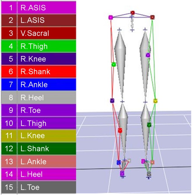
전방 점프 착지 동작의 정확한 분석을 위해 8대의 동작분석 카메라(6 Eagle Camera, 2 Raptor-E Camera, Motion Analysis Corp., USA)가 좌표 기준점과 대상자 동작 범위를 포함할 수 있도록 공간좌표 설정을 위해 영점 조절(calibration)을 실시하였다. 진행방향을 Y축, 좌우방향을 X축, 수직방향을 Z축으로 설정하였으며, 대상자의 3차원적 자료 수집을 위해 하지에 10 mm 직경의 반사 마커 19개를 Helen Hayes Marker Set (Kadaba et al., 1989)에 의거하여 부착하였다. 반사 마커 부착 후 해부학적 정적 자세를 촬영하고 무릎과 발목관절에 부착된 내측 마커 4개를 제거한 뒤 동작을 수행하였다(Figure 1).

전방 점프 착지 동작을 수행하기 위해 Sell (2012)의 연구를 바탕으로 대상자와 지면반력기 중심까지의 수평거리는 대상자 신장의 40%로 설정하였으며, 충분한 수직 점프가 이루어질 수 있도록 대상자와 지면반력기 중간 지점에 30 cm 높이의 장애물을 설치하였다. 설치 후 대상자는 시작 지점에서 맨발인 상태로 양 발로 점프하여 장애물을 넘은 뒤 전방에 위치한 지면반력기에 한 발로 착지하였다. 동작 수행 시 상지 움직임의 영향을 최소화하기 위해 모든 대상자는 양 손을 골반에 부착한 마커를 가리지 않도록 주의하여 장골능에 고정시켰다.
전방 점프 착지 동작을 수행하기 전 부상을 예방하기 위해 최소 30분의 준비 운동을 충분히 실시하였으며, 양 발로 전방 점프 후 한 발로 지면반력기 중앙에 착지하였다. 이때 정상 집단은 비우성 하지 (non-dominant leg)로 착지하였으며, 만성 발목 불안정성 집단의 경우 발목 불안정성이 있는 쪽 하지로 착지를 수행하였다. 정상 집단의 비우성 하지는 공을 찰 때 주로 사용하는 발을 우성 하지(dominant leg)로 정의하고 반대쪽 발을 비우성 하지로 정의하였다. 두 집단 모두 5회 이상 동작을 수행하였으며, 동작 수행 중 착지 시 발이 지면반력기 밖으로 벗어나거나 착지 후 발이 지면으로부터 다시 떨어지는 경우, 장골능에 고정한 손이 떨어지는 경우, 넘어지는 경우, 착지 후 발이 지면반력기 내에 부분적으로 걸치는 경우는 실패로 간주하였다. 정상 집단의 경우 우성 하지로 착지하는 경우 및 만성 발목 불안정성 집단에서 발목 불안정성이 나타나지 않는 하지로 착지하는 경우는 실패로 간주하였다. 실패 조건에 해당되는 동작을 제외하고 가장 안정적인 착지 3회의 측정치를 평균값으로 산출하여 분석에 사용하였다.
3. 자료처리 및 분석
전방 점프 착지 동작의 운동학적 변인 분석을 위해 총 8대의 동작분석 카메라를 통해 120 frame/sec로 수집하였으며, 수집된 자료는 Cortex 5.0 (Motion Analysis Corp., USA)를 이용하여 분석하였다. 2차원 상의 평면 데이터는 NLT (non-linear transformation) 방식에 의해 3차원 데이터로 변환하였으며, 오차 제거를 위해 저역통과 필터링(Butterworth filter)를 통해 스무딩하였다.
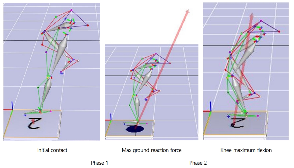
지면반력 데이터 수집은 1대의 지면반력기(AMTI inc., USA)를 사용하였으며, 초당 1,200 Hz로 샘플링하여 분석하였다. 동작분석 및 지면반력 데이터는 아날로그-디지털 컨버터(A/D Converter, NI USB-6218, National Instruments, Hungary)를 통해 동기화한 후 측정하였다. 지면으로부터 발생하는 수직지면반력 데이터는 각 대상자의 몸무게(body weight, BW)로 나누어 정규화한 후 비교, 분석하였다. 모든 결과 변인은 (Figure 2)와 같이 초기접지(initial contact, IC), 최대수직지면반력(max ground reaction force, mGRF), 최대무릎굴곡(knee maximum flexion, KMF) 시점으로 구분하여 분석하였으며, 초기접지 시점에서 최대수직지면반력 시점까지를 구간 1 (phase 1), 최대수직지면반력 시점에서 최대무릎굴곡 시점을 구간 2 (phase 2)로 정의하였다.

(1) 하지관절 각도의 정의
전방 점프 후 착지 동작에 대한 하지관절의 시상면, 관상면, 수평면에서의 각도 정의는 (Table 2)과 같다. 엉덩관절 각도는 골반에 대한 허벅지 분절의 움직임, 무릎관절 각도는 허벅지 분절에 대한 정강이 분절의 움직임을 반영하며, 발목관절 각도는 정강이 분절에 대한 발 분절의 움직임을 반영한다. 시상면 움직임은 내외축을 기준으로 나타나며, 엉덩관절과 무릎관절은 굴곡/신전 움직임을 의미하고 발목관절은 족저굴곡/배측굴곡 움직임을 의미한다. 관상면 움직임은 전후축을 기준으로 나타나는 움직임으로 엉덩관절은 허벅지 분절의 외전/내전, 무릎관절은 정강이 분절의 내반/외반, 마지막으로 발목관절은 후족부의 외번/내번 움직임으로 정의한다. 수평면 움직임은 수직축을 기준으로 나타나는 내회전 및 외회전 움직임을 의미한다.
|
Plane |
Joint |
Anatomical |
Anatomical |
|
Sagittal |
Hip |
Flexion |
Extension |
|
Knee |
Flexion |
Extension |
|
|
Ankle |
Plantar flexion |
Dorsiflexion |
|
|
Frontal |
Hip |
Abduction |
Adduction |
|
Knee |
Valgus |
Varus |
|
|
Ankle |
Eversion |
Inversion |
|
|
Transverse |
Hip |
Internal rotation |
External rotation |
|
Knee |
Internal rotation |
External rotation |
|
|
Ankle |
External rotation |
Internal rotation |
4. 통계처리
본 연구의 통계처리는 Windows용 SPSS 26.0 (IBM, USA)을 이용하였고, 측정된 모든 변수에 대해 평균 및 표준편차를 산출하였다. 정상 집단과 만성 발목 불안정성 집단의 전방 점프 후 착지 동작을 비교, 분석하기 위해 독립표본 t 검정(independent t-test)을 실시하였으며, 모든 통계적인 유의수준은 p <.05로 설정하였다.
1. 운동학적 변인
(1) 엉덩관절 각도
전방 점프 후 착지 시 두 집단 간 엉덩관절 각도를 분석한 결과, 초기접지 시점의 시상면 및 관상면 각도에서 유의한 차이가 있는 것으로 나타났으며, CAI 집단은 정상 집단과 비교하여 굴곡 각도(p=0.000)와 외전 각도(p=0.044)가 유의하게 작았다. 최대수직지면반력(p=0.000) 및 무릎최대굴곡 시점(p=0.000)의 경우 CAI 집단이 정상 집단에 비해 굴곡 각도가 유의하게 작은 것으로 나타났다. 이 외에 다른 시점 및 엉덩관절의 움직임에서는 유의한 차이가 나타나지 않았다(Table 3).
|
Event |
Plane |
CON |
CAI |
p-value |
|
IC |
Sagittal |
41.8±6.5 |
27.2±8.2 |
.000 |
|
Frontal |
8.2±5.4 |
4.9±4.6 |
.044 |
|
|
Transverse |
-3.7±11.5 |
-5.9±7.8 |
.479 |
|
|
mGRF |
Sagittal |
45.6±7.3 |
27.8±8.1 |
.000 |
|
Frontal |
3.9±6.6 |
4.7±5.6 |
.682 |
|
|
Transverse |
5.1±9.5 |
0.5±7.8 |
.100 |
|
|
MKF |
Sagittal |
48.5±7.9 |
35.0±9.2 |
.000 |
|
Frontal |
0.0±7.6 |
-2.7±5.8 |
.218 |
|
|
Transverse |
9.9±8.8 |
6.2±7.8 |
.204 |
|
|
Note. (+) value: Flexion, abduction, internal
rotation, (-) value: Exten- sion,
adduction, external rotation, IC: initial contact, mGRF: max ground reaction
force, MKF: maximum knee flexion, CON: control group, CAI: chronic ankle instability group, Data are mean ± standard deviation |
||||
(2) 무릎관절 각도
무릎관절의 시상면 움직임을 분석한 결과, 초기접지 시점에서 CAI 집단은 정상 집단보다 굴곡 각도가 작은 것으로 나타났으며(p=0.041), 관상면 움직임의 경우 정상 집단은 내반 움직임이 나타난 반면 CAI 집단은 외반 움직임으로 유의한 차이가 나타났다(p=0.000). 최대수직지면반력 시점은 초기접지 시점과 마찬가지로 시상면 및 관상면 움직임에서 유의한 차이가 관찰되었다. 구체적으로 시상면 움직임에서 CAI 집단은 정상 집단보다 굴곡 각도가 작았고(p=0.000), 관상면 움직임의 경우 CAI 집단의 내반 각도가 유의하게 작은 것으로 나타났다(p=0.000). 무릎최대굴곡 시점에서는 두 집단 간 관상면 움직임에서 유의한 차이가 나타났으며(p=0.000), CAI 집단은 정상 집단보다 내반 각도가 작았다. 이 외에 수평면 움직임에서는 모든 시점에서 유의한 차이는 나타나지 않았다(Table 4).
|
Event |
Plane |
CON |
CAI |
p-value |
|
IC |
Sagittal |
17.4±7.7 |
13.3±3.6 |
.041 |
|
Frontal |
-5.9±3.8 |
0.8±3.8 |
.000 |
|
|
Transverse |
-15.4±10.1 |
-17.2±9.1 |
.572 |
|
|
mGRF |
Sagittal |
33.3±6.4 |
26.0±5.6 |
.000 |
|
Frontal |
-11.0±4.5 |
-1.9±5.3 |
.000 |
|
|
Transverse |
-15.3±10.6 |
-13.4±6.2 |
.495 |
|
|
MKF |
Sagittal |
42.3±7.4 |
42.3±7.4 |
.999 |
|
Frontal |
-13.7±4.6 |
-4.5±5.8 |
.000 |
|
|
Transverse |
-13.2±10.5 |
-12.7±7.4 |
.855 |
|
|
Note. (+) value: Flexion,
valgus, internal rotation, (-)
value: Extension, varus,
external rotation, IC: initial contact, mGRF: max ground reaction force, MKF: maximum knee flexion, CON: control group, CAI:
chronic ankle instability group, Data are mean ± standard
deviation |
||||
(3) 발목관절 각도
초기접지 시점의 경우 발목관절의 모든 움직임에서 유의한 차이가 나타났다. 시상면 움직임에서 CAI 집단은 정상 집단에 비해 족저굴곡 각도가 컸으며(p=0.001), 관상면 움직임의 경우 CAI 집단이 내번 각도가 유의하게 큰 것으로 나타났다(p=0.003). 수평면 움직임에서는 CAI 집단은 정상 집단보다 내회전 각도가 유의하게 컸다(p=0.000). 최대수직지면반력 시점의 관상면 및 수평면 움직임에서 두 집단 간 유의한 차이가 나타났다. 먼저 관상면 움직임에서 CAI 집단이 내번 각도가 유의하게 컸으며(p=0.002), 수평면 움직임에서는 CAI 집단의 경우 내회전 움직임, 정상 집단은 외회전 움직임이 나타났다(p= 0.021). 마지막으로 무릎최대굴곡 시점의 경우 관상면 움직임에서만 유의한 차이가 관찰되었으며(p=0.043), 다른 시점과 마찬가지로 CAI 집단의 내번 각도가 유의하게 큰 것으로 나타났다(Table 5).
|
Event |
Plane |
CON |
CAI |
p-value |
|
IC |
Sagittal |
28.4±8.4 |
44.2±18.6 |
.001 |
|
Frontal |
-3.7±6.0 |
-9.4±5.3 |
.003 |
|
|
Transverse |
-4.2±8.3 |
-14.4±4.7 |
.000 |
|
|
mGRF |
Sagittal |
11.4±6.2 |
18.1±19.2 |
.140 |
|
Frontal |
-9.3±5.7 |
-16.5±7.9 |
.002 |
|
|
Transverse |
0.5±9.5 |
-5.2±4.4 |
.021 |
|
|
MKF |
Sagittal |
3.6±5.5 |
1.7±19.8 |
.679 |
|
Frontal |
-12.8±6.0 |
-17.7±8.5 |
.043 |
|
|
Transverse |
3.9±9.4 |
3.8±6.6 |
.965 |
|
|
Note. (+) value: Plantar flexion, eversion, external rotation, (-) value: Dorsiflexion, inversion, internal
rotation, IC: initial contact, mGRF: max ground
reaction force, MKF: maximum knee flexion, CON: control group, CAI: chronic ankle
instability group, Data are mean ± standard deviation |
||||
2. 운동역학적 변인
(1) 수직지면반력
전방 점프 착지 후 두 집단 간의 수직지면반력 비교 결과 초기접지 및 최대수직지면반력 시점에서 유의한 차이가 나타났다. 초기접지 시점에서 CAI 집단은 정상 집단보다 높은 수직지면반력이 나타났으며(p=0.000), 최대수직지면반력 시점 또한 CAI 집단이 정상 집단에 비해 유의하게 높은 수직지면반력이 나타났다(p=0.000) (Table 5).
(2) 압력중심
전방 점프 착지 후 두 집단 간의 COP X 및 COP Y 비교 결과 COP X은 최대수직지면반력 시점에서 CAI 집단은 정상 집단보다 유의하게 압력중심이 뒤로 이동하였다(p=0.032). COP Y는 모든 시점에서 두 집단 간 유의한 차이가 나타났으며(p<0.05), 모든 시점에서 정상 집단은 내측으로 이동한 압력중심이 나타난 반면, CAI 집단은 외측으로 이동한 압력중심이 나타났다(Table 6).
|
Event |
Variables |
CON |
CAI |
p-value |
|
IC |
GRF (N) |
0.12±0.13 |
0.50±0.34 |
.000 |
|
COP X (mm) |
17.89±70.24 |
23.93±43.08 |
.745 |
|
|
COP Y (mm) |
53.88±73.09 |
-4.53±41.83 |
.004 |
|
|
mGRF |
GRF (N) |
2.14±0.41 |
3.57±0.48 |
.000 |
|
COP X (mm) |
-2.33±36.55 |
-32.58±48.53 |
.032 |
|
|
COP Y (mm) |
17.65±31.09 |
-12.17±38.29 |
.010 |
|
|
MKF |
GRF (N) |
1.70±0.32 |
1.63±0.32 |
.466 |
|
COP X (mm) |
0.65±53.90 |
-7.68±50.74 |
.618 |
|
|
COP Y (mm) |
17.82±39.10 |
-13.50±37.58 |
.014 |
|
|
Note. (+) value: Front, medial
direction, (-) value:
Posterior, lateral direction, IC: initial contact, mGRF: max ground reaction
force, MKF: maximum knee flexion, CON: control group, CAI: chronic ankle in- stability
group, Data are mean ± standard deviation |
||||
이 연구는 전방 점프 착지 시 만성 발목 불안정성 유무가 자세 조절에 어떠한 영향을 미치는지 조사하기 위해 하지관절의 운동학적 및 운동역학적 분석을 통해 만성 발목 불안정성 집단과 정상 집단을 구분하여 착지 전략에 차이가 나타나는지 규명하고자 하였다.
이전 연구에서 만성 발목 불안정성이 있는 경우 이에 따른 보상작용으로 지면으로부터 발생하는 충격을 감소시키고 안정적인 착지를 유도하기 위해 엉덩관절을 굴곡시키는 전략을 선택한다고 하였으나(Lin et al., 2019; Yu et al., 2021), 다른 연구에서는 엉덩관절은 착지 동작 시 정상 집단과 큰 차이가 나타나지 않는다고 보고하였다(De Ridder, Willems, Vanrenterghem, Robinson & Roosen, 2015; Gribble & Robinson, 2009; Terada, Pietrosimone & Gribble, 2014). 그러나 연구 결과 모든 시점에서 만성 발목 불안정성 집단은 정상 집단보다 유의하게 작은 엉덩관절 굴곡 각도가 나타났다. 이러한 결과의 차이는 만성 발목 불안정성 집단에서 엉덩관절의 굴곡 각도가 크다고 보고한 연구에서는 대부분 드롭랜딩과 같은 자유 낙하(Doherty et al., 2015) 또는 수직 점프를 수행하였으며(Terada et al., 2014), 동작 형태에 기인한 것으로 사료된다. 만성 발목 불안정성 집단은 엉덩관절 굴곡 각도와 함께 무릎관절 각도가 정상 집단에 비해 유의하게 작은 것으로 나타났는데 무릎 및 엉덩관절의 굴곡 각도가 증가할수록 무릎관절에 전달되는 부하가 감소하며, 굴곡 각도가 감소할수록 햄스트링과 전방십자인대가 수평에 가까워져 경골의 전방 전위를 제한하는 햄스트링의 능력을 제한하게 된다(Alentorn-Geli et al., 2009). 이는 전방십자인대와 같은 연부 조직에 부하를 증가시켜 부상 위험을 증가시킬 수 있다는 관점에서 중요한 결과이다(Bakker et al., 2016; Yu, Lin & Garrett, 2006).
만성 발목 불안정성 집단의 무릎관절 관상면 움직임 또한 무릎관절 부상 위험 증가와 연관될 수 있는 결과가 나타났다. 구체적으로 만성 발목 불안정성 집단은 초기접지 시점에서 정상 집단과 달리 외반 움직임이 나타나고 최대수직지면반력 시점에서 작은 내반 각도가 나타났다. 특히 무릎관절의 외반 움직임은 최근 외측 발목 염좌의 예측 인자로서 보고될 뿐만 아니라(Mineta et al., 2021) 전방십자인대 손상의 중요한 요인이며, 실제 스포츠 상황에서 착지 동작 시 전방십자인대 손상 사례를 분석한 결과 모든 대상자가 초기접지 시점에서 40 ms 이내에 외반 움직임이 나타났다고 보고하였다(Koga et al., 2010). 또한 이전 만성 발목 불안정성의 양 발 착지 시 정상 집단에 비해 높은 외반 각도가 나타나 이는 전방십자인대 부상 위험이 증가할 수 있다고 보고한 연구 결과와 일치한다(Jeon & Park, 2021). 이러한 결과는 만성 발목 불안정성 집단에서 무릎 및 엉덩관절의 시상면 움직임 감소와 무릎관절의 외반 움직임이 나타나 비접촉성 무릎 부상 위험이 높은 착지 동작을 수행하는 것을 시사한다.
하지관절은 착지 시 발생하는 충격을 최소화하기 위해 시상면 움직임을 사용하는데 이때 발목관절의 시상면 움직임의 관절 가동 범위는 지면반력 흡수에 중요한 역할을 담당하고, 발목관절의 배측굴곡 각도가 감소할수록 지면반력은 증가하게 된다(Self & Paine, 2001). 이 연구에서 발목관절의 족저굴곡 각도는 초기접지 시점에서 만성 발목 불안정성 집단이 정상 집단보다 유의하게 크게 나타나고 수직지면반력은 초기접지 및 최대수직지면반력 시점에서 정상 집단보다 높았다. 이는 무릎 및 엉덩관절의 굴곡 각도를 통해 충격을 흡수한 정상 집단과는 달리 만성 발목 불안정성은 다른 하지관절에서 굴곡 각도를 증가시키지 못하고 발목관절이 족저굴곡 된 상태로 착지하여 충격을 흡수하는 전략을 사용한 것으로 사료된다. 착지 시 후족부가 먼저 닿는 형태보다 전족부를 통해 수행할 경우 수직지면반력이 감소하게 되는데 이는 발목관절의 시상면 움직임의 가동 범위를 증가시키기 때문이다. 그러나 이전 연구와 마찬가지로 만성 발목 불안정성 집단은 정상 집단보다 수직지면반력이 유의하게 높았다(Herb et al., 2018; Kim, Son, Seeley & Hopkins, 2019). 이를 토대로 만성 발목 불안정성 집단은 다른 하지관절의 굴곡 각도 감소를 보상하기 위해 족저굴곡 각도가 높은 상태로 착지하였으나, 수직지면반력을 충분히 흡수하지 못한 것으로 판단된다.
만성 발목 불안정성 환자의 큰 족저굴곡 각도 및 내번 각도의 증가는 발목 염좌 부상 위험의 주요 원인으로 알려져 있다(Panagiotakis et al., 2017). 이전 연구와 마찬가지로 연구 결과, 만성 발목 불안정성 집단은 모든 시점에서 정상 집단보다 큰 내번 각도가 나타났다. 특히, 초기접지 시점의 경우 족저굴곡 및 내번 각도가 크게 나타났는데 이는 발목 염좌 재발 위험을 증가시킬 수 있음을 시사한다. 또한 큰 내번 각도로 인해 만성 발목 불안정성 집단은 외측으로 이동한 압력중심이 나타났으며, 이는 발이 지면에 접촉한 상태에서 발목관절의 내번으로 인해 압력중심이 외측에 위치한다는 선행연구(Nawata, Nishihara, Hayashi & Teshima, 2005; Pope et al., 2011)와 일치하여 만성 발목 불안정성 집단의 자세 조절 능력의 감소를 나타냈다. 외측으로 이동한 압력중심은 외측 발목 염좌의 재발 뿐만 아니라 내측 거골 연골에 가해지는 부하를 증가시켜 골관절염과 같은 장기적인 문제를 초래(Wenning, Lange, Paul, Gollhofer & Gehring, 2019)하기 때문에 발목관절의 운동학적 변화 및 자세 조절 능력 감소를 개선하여 외측 발목 염좌 재발 위험성을 감소시키는 것이 중요하다.
이전 만성 발목 불안정성을 대상으로 한 선행연구에서는 지속적인 발목 염좌로 인해 착지 시 발목관절 내번 각도 증가, 무릎관절 굴곡 각도 감소, 엉덩관절 굴곡 각도를 증가시켜 정상 집단과 달리 엉덩관절을 사용한 착지 패턴의 변화가 나타난다고 보고하였다(Lin et al., 2019; Yu et al., 2021). 그러나 연구 결과 만성 발목 불안정성 집단은 전방 점프 착지 시 엉덩 및 무릎관절의 굴곡 각도가 감소하고 무릎관절의 외반 각도가 증가하였으며, 발목관절의 내번 각도 증가와 함께 수직지면반력이 큰 것으로 나타났다. 이러한 결과를 토대로 만성 발목 불안정성 집단은 정상 집단과 다른 착지 전략이 나타나는 것을 확인할 수 있었으며, 이와 더불어 만성 발목 불안정성 집단에서 나타난 하지관절의 운동학적 및 운동역학적 변화는 외측 발목 염좌 위험을 증가시킬 뿐만 아니라 전방십자인대 손상과 같은 비접촉성 무릎 부상 요인이 될 수 있음을 시사한다.
이 연구는 전방 점프 착지 시 만성 발목 불안정성 유무가 자세 조절에 어떠한 영향을 미치는지 조사하기 위해 하지관절의 운동학적 및 운동역학적 분석을 조사하였다. 결과에 따르면 전방 점프 후 착지 시 만성 발목 불안정성 집단은 정상 집단보다 모든 시점에서 발목관절의 큰 내번 각도와 외측으로 이동한 압력중심이 나타났으며, 초기접지 및 최대수직지면반력 시점에서 엉덩 및 무릎관절의 굴곡 각도가 작고, 무릎관절의 외반 움직임 및 최대수직지면반력이 유의하게 큰 것으로 나타났다. 이를 통해 전방 점프 후 착지 시 만성 발목 불안정성 집단은 하지관절의 운동학적 변화로 인해 충격 흡수를 적절히 수행하지 못하고 외측 발목 염좌 재발 위험이 높은 형태의 착지를 수행하였으며, 이는 결국 인접한 관절의 부하 증가 및 연부조직 손상의 부상 요인이 될 수 있으므로 이를 예방하기 위한 추가적인 연구가 필요할 것이다.
References
1. Alentorn-Geli, E., Myer, G. D., Silvers, H. J., Samitier, G., Romero, D., Lázaro-Haro, C. & Cugat, R. (2009). Prevention of non-contact anterior cruciate ligament injuries in soccer players. Part 1: Mech- anisms of injury and underlying risk factors. Knee Surgery, Sports Traumatology, Arthroscopy, 17(7), 705-729.
Google Scholar
2. Ali, N., Robertson, D. G. E. & Rouhi, G. (2014). Sagittal plane body kinematics and kinetics during single-leg landing from increasing vertical heights and horizontal distances: Implications for risk of non-contact ACL injury. The Knee, 21(1), 38-46.
Google Scholar
3. Anandacoomarasamy, A. & Barnsley, L. (2005). Long term outcomes of inversion ankle injuries. British Journal of Sports Medicine, 39(3), e14.
Google Scholar
4. Ardakani, M. K., Wikstrom, E. A., Minoonejad, H., Rajabi, R. & Sharifnezhad, A. (2019). Hop-stabilization training and landing bio- mechanics in athletes with chronic ankle instability: a randomized controlled trial. Journal of Athletic Training, 54(12), 1296-1303.
Google Scholar
5. Bakker, R., Tomescu, S., Brenneman, E., Hangalur, G., Laing, A. & Chandrashekar, N. (2016). Effect of sagittal plane mechanics on ACL strain during jump landing. Journal of Orthopaedic Research, 34(9), 1636-1644.
Google Scholar
6. Brown, C., Bowser, B. & Simpson, K. J. (2012). Movement variability during single leg jump landings in individuals with and without chronic ankle instability. Clinical Biomechanics, 27(1), 52-63.
Google Scholar
7. Brown, C. N., Padua, D. A., Marshall, S. W. & Guskiewicz, K. M. (2009). Variability of motion in individuals with mechanical or functional ankle instability during a stop jump maneuver. Clinical Biomechanics, 24(9), 762-768.
Google Scholar
8. De Ridder, R., Willems, T., Vanrenterghem, J., Robinson, M. A. & Roosen, P. (2015). Lower limb landing biomechanics in subjects with chronic ankle instability. Medicine and Science in Sports and Exercise, 47(6), 1225-1231.
Google Scholar
9. Delahunt, E., Monaghan, K. & Caulfield, B. (2006). Changes in lower limb kinematics, kinetics, and muscle activity in subjects with functional instability of the ankle joint during a single leg drop jump. Journal of Orthopaedic Research, 24(10), 1991-2000.
Google Scholar
10. Doherty, C., Bleakley, C., Hertel, J., Caulfield, B., Ryan, J., Sweeney, K., Paterson, M. & Delahunt, E. (2015). Coordination and symmetry patterns during the drop vertical jump, 6-months after first-time lateral ankle sprain. Journal of Orthopaedic Research, 33(10), 1537-1544.
Google Scholar
11. Fong, D., Hong, Y., Chan, L. K., Yung, P. & Chan, K. M. (2007). A systematic review on ankle injury and ankle sprain in sports. Sports Medicine, 37(1), 73-94.
Google Scholar
12. Gribble, P. A. & Robinson, R. H. (2009). Alterations in knee kinematics and dynamic stability associated with chronic ankle instability. Journal of Athletic Training, 44(4), 350-355.
Google Scholar
13. Herb, C. C., Grossman, K., Feger, M. A., Donovan, L. & Hertel, J. (2018). Lower extremity biomechanics during a drop-vertical jump in participants with or without chronic ankle instability. Journal of Athletic Training, 53(4), 364-371.
Google Scholar
14. Hertel, J. (2002). Functional anatomy, pathomechanics, and patho- physiology of lateral ankle instability. Journal of Athletic Training, 37(4), 364-375.
Google Scholar
15. Hertel, J. & Corbett, R. O. (2019). An updated model of chronic ankle instability. Journal of Athletic Training, 54(6), 572-588.
Google Scholar
16. Hiller, C. E., Nightingale, E. J., Lin, C. W. C., Coughlan, G. F., Caulfield, B. & Delahunt, E. (2011). Characteristics of people with recurrent ankle sprains: a systematic review with meta-analysis. British Journal of Sports Medicine, 45(8), 660-672.
Google Scholar
17. Hintermann, B., Boss, A. & Schäfer, D. (2002). Arthroscopic findings in patients with chronic ankle instability. The American Journal of Sports Medicine, 30(3), 402-409.
Google Scholar
18. Hoch, M. C. & McKeon, P. O. (2014). Peroneal reaction time after ankle sprain: a systematic review and meta-analysis. Medicine and Science in Sports and Exercise, 46(3), 546-556.
Google Scholar
19. Hootman, J. M., Dick, R. & Agel, J. (2007). Epidemiology of collegiate injuries for 15 sports: summary and recommendations for injury prevention initiatives. Journal of Athletic Training, 42(2), 311-319.
Google Scholar
20. Jeon, K. & Park, J. (2021). Biomechanical Characteristic on Lower Extremity with or without Chronic Ankle Instability during Double Leg Drop Landing. Korean Journal of Sport Biomechanics, 31(2), 113-118.
Google Scholar
21. Kadaba, M., Ramakrishnan, H., Wootten, M., Gainey, J., Gorton, G. & Cochran, G. (1989). Repeatability of kinematic, kinetic, and electro- myographic data in normal adult gait. Journal of Orthopaedic Research, 7(6), 849-860.
Google Scholar
22. Kim, H., Son, S. J., Seeley, M. K. & Hopkins, J. T. (2019). Altered move- ment biomechanics in chronic ankle instability, coper, and control groups: energy absorption and distribution implications. Journal of Athletic Training, 54(6), 708-717.
Google Scholar
23. Koga, H., Nakamae, A., Shima, Y., Iwasa, J., Myklebust, G., Engebretsen, L., Bahr. R. & Krosshaug, T. (2010). Mechanisms for noncontact anterior cruciate ligament injuries: knee joint kinematics in 10 injury situations from female team handball and basketball. The American journal of Sports Medicine, 38(11), 2218-2225.
Google Scholar
24. Kotsifaki, A., Korakakis, V., Graham-Smith, P., Sideris, V. & Whiteley, R. (2021). Vertical and horizontal hop performance: contributions of the hip, knee, and ankle. Sports Health, 13(2), 128-135.
Google Scholar
25. Kunugi, S., Masunari, A., Yoshida, N. & Miyakawa, S. (2017). Postural stability and lower leg muscle activity during a diagonal single-leg landing differs in male collegiate soccer players with and without functional ankle instability. The Journal of Physical Fitness and Sports Medicine, 6(4), 257-265.
Google Scholar
26. Lin, J. Z., Lin, Y. A. & Lee, H. J. (2019). Are landing biomechanics altered in elite athletes with chronic ankle instability. Journal of Sports Science & Medicine, 18(4), 653-662.
Google Scholar
27. Marshall, A. N., Hertel, J., Hart, J. M., Russell, S. & Saliba, S. A. (2020). Visual biofeedback and changes in lower extremity kinematics in individuals with medial knee displacement. Journal of Athletic Training, 55(3), 255-264.
Google Scholar
28. McNitt-Gray, J. L. (1991). Kinematics and impulse characteristics of drop landings from three heights. Journal of Applied Biomechanics, 7(2), 201-224.
Google Scholar
29. Mineta, S., Inami, T., Hoshiba, T., Higashihara, A., Kumai, T., Torii, S. & Hirose, N. (2021). Greater knee varus angle and pelvic internal rotation after landing are predictive factors of a non-contact lateral ankle sprain. Physical Therapy in Sport, 50, 59-64.
Google Scholar
30. Mitchell, A., Dyson, R., Hale, T. & Abraham, C. (2008). Biomechanics of ankle instability. Part 2: Postural sway-reaction time relationship. Medicine and Science in Sports and Exercise, 40(8), 1522-1528.
Google Scholar
31. Montalvo, A. M., Schneider, D. K., Webster, K. E., Yut, L., Galloway, M. T., Heidt Jr, R. S. ... & Parikh, S. N. (2019). Anterior cruciate ligament injury risk in sport: a systematic review and meta-analysis of injury incidence by sex and sport classification. Journal of Athletic Training, 54(5), 472-482.
Google Scholar
32. Nawata, K., Nishihara, S., Hayashi, I. & Teshima, R. (2005). Plantar pres- sure distribution during gait in athletes with functional instability of the ankle joint: preliminary report. Journal of Orthopaedic Science, 10(3), 298-301.
Google Scholar
33. Panagiotakis, E., Mok, K. M., Fong, D. T. P. & Bull, A. M. (2017). Bio- mechanical analysis of ankle ligamentous sprain injury cases from televised basketball games: understanding when, how and why ligament failure occurs. Journal of Science and Medicine in Sport, 20(12), 1057-1061.
Google Scholar
34. Pope, M., Chinn, L., Mullineaux, D., McKeon, P. O., Drewes, L. & Hertel, J. (2011). Spatial postural control alterations with chronic ankle instability. Gait & Posture, 34(2), 154-158.
Google Scholar
35. Self, B. P. & Paine, D. (2001). Ankle biomechanics during four landing techniques. Medicine and Science in Sports and Exercise, 33(8), 1338-1344.
Google Scholar
36. Sell, T. C. (2012). An examination, correlation, and comparison of static and dynamic measures of postural stability in healthy, physically active adults. Physical Therapy in Sport, 13(2), 80-86.
Google Scholar
37. Struijs, P. A. & Kerkhoffs, G. M. (2010). Ankle sprain. BMJ Clin Evid, 2010.
Google Scholar
38. Swenson, D. M., Collins, C. L., Fields, S. K. & Comstock, R. D. (2013). Epidemiology of US high school sports-related ligamentous ankle injuries, 2005/06-2010/11. Clinical Journal of Sport Medicine: Offi- cial Journal of the Canadian Academy of Sport Medicine, 23(3), 190-196.
Google Scholar
39. Tanen, L., Docherty, C. L., Van Der Pol, B., Simon, J. & Schrader, J. (2014). Prevalence of chronic ankle instability in high school and division I athletes. Foot & Ankle Specialist, 7(1), 37-44.
Google Scholar
40. Terada, M., Pfile, K. R., Pietrosimone, B. G. & Gribble, P. A. (2013). Effects of chronic ankle instability on energy dissipation in the lower extremity. Medicine and Science in Sports and Exercise, 45(11), 2120-2128.
Google Scholar
41. Terada, M., Pietrosimone, B. & Gribble, P. A. (2014). Individuals with chronic ankle instability exhibit altered landing knee kinematics: potential link with the mechanism of loading for the anterior cruciate ligament. Clinical Biomechanics, 29(10), 1125-1130.
Google Scholar
42. Theisen, A. & Day, J. (2019). Chronic ankle instability leads to lower extremity kinematic changes during landing tasks: a systematic review. International Journal of Exercise Science, 12(1), 24-33.
Google Scholar
43. Van Der Does, H., Brink, M., Benjaminse, A., Visscher, C. & Lemmink, K. (2016). Jump landing characteristics predict lower extremity injuries in indoor team sports. International Journal of Sports Medicine, 37(03), 251-256.
Google Scholar
44. Wenning, M., Lange, T., Paul, J., Gollhofer, A. & Gehring, D. (2019). Assessing mechanical ankle instability via functional 3D stress-MRI–a pilot study. Clinical Biomechanics, 70, 107-114.
Google Scholar
45. Wernli, K., Ng, L., Phan, X., Davey, P. & Grisbrook, T. (2016). The relation- ship between landing sound, vertical ground reaction force, and kinematics of the lower limb during drop landings in healthy men. Journal of Orthopaedic & Sports Physical Therapy, 46(3), 194-199.
Google Scholar
46. Yu, B., Lin, C. F. & Garrett, W. E. (2006). Lower extremity biomechanics during the landing of a stop-jump task. Clinical Biomechanics, 21(3), 297-305.
Google Scholar
47. Yu, P., Mei, Q., Xiang, L., Fernandez, J. & Gu, Y. (2021). Differences in the locomotion biomechanics and dynamic postural control between individuals with chronic ankle instability and copers: a systematic review. Sports Biomechanics, 1-19.
Google Scholar