Open Access, Peer-reviewed
eISSN 2093-9752
Open Access, Peer-reviewed
eISSN 2093-9752
Sang-Kyoon Park
Seungbum Koo
Suk-Hoon Yoon
Sangheon Park
Yongcheol Kim
Ji-Seon Ryu
http://dx.doi.org/10.5103/KJSB.2018.28.2.101 Epub 2018 July 13
Abstract
Objective: The aim of this study was to investigate gender differences in ground reaction force (GRF) components among different speeds of running.
Method: Twenty men (age=22.4±1.6 years, mass=73.4±8.4 kg, height=176.2±5.6 cm) and twenty women (age=20.7±1.2 years, mass=55.0±8.2 kg, height=163.9±5.3 cm) participated in this study. All participants were asked to run on an instrumented dual belt treadmill (Bertec, USA) at 8, 12, and 16 km/h for 3 min, after warming up. GRF data were collected from 30 strides while they were running. Hypotheses were tested using one-way ANOVA, and level of significance was set at p-value <.05.
Results: The time to passive peaks was significantly earlier in women than in men at three different running speeds (p<.05). Further, the impact loading rates were significantly greater in women than in men at three different running speeds (p<.05). Moreover, the propulsive peak at 8 km/h, which is the slowest running speed, was significantly greater in women than in men (p<.05), and the vertical impulse at 16 km/h, which is the fastest running speed, was significantly greater in men than in women (p<.05). The absolute anteroposterior impulse at 8 km/h was significantly greater in women than in men (p<.05). In addition, as the running speed increased, impact peak, active peak, impact loading rate, breaking peak, propulsive peak, and anteroposterior impulse were significantly increased, but vertical impulse was significantly decreased (p<.05).
Conclusion: The impact loading rate is greater in women than in men regardless of different running speeds. Therefore, female runners might be exposed to the risk of potential injuries related to the bone and ligament. Moreover, increased running speeds could lead to higher possibility of running injuries.
Keywords
Running Gender Speed GRF Loading rate
Running is one of the most popular exercises that is performed throughout the lifetime as it has relatively fewer spatial restrictions compared with other exercises. Running regularly has many physiological benefits but can also cause chronic overuse injuries. About 20~80% of all skeletal injuries are reported to be due to running (Bredeweg & Buist, 2011; Lun, Meeuwisse, Stergiou, & Stefanyshyn, 2004; Taunton et al., 2003; van Gent et al., 2007). Running injuries are caused by various factors. They are affected by internal factors such as dynamic abnormalities, previous injuries, gender, and body mass index and by external factors such as training frequency, intensity, and duration; muscle strength; flexibility; and shoes (Taunton et al., 2003; van Mechelen, 1992).
Of the factors that affect running injuries, gender difference is an important topic of research. Robinson and Nee (2007) claimed that female athletes are at increased risk of chronic injuries due to running compared with male athletes of the same age. Geraci and Brown (2005) and Taunton et al. (2002) reported that the risk of running injuries such as patellofemoral pain syndrome, iliotibial band syndrome, and injuries of the gluteus medius muscle is two times higher among women than among men. Lopes, Hespanhol, Yeong, and Costa (2012) reported that patellofemoral pain syndrome is the most common injury that occurs during running and that its incidence and prevalence are higher among women. Taunton et al. (2003) and van Gent et al. (2007) further characterized the different sites of injury between men and women by claiming that injuries of the calves and feet due to running are more common among men, whereas those of the hip are more common among women.
In addition to research aimed at understanding the properties of running injuries in men and women, various approaches have been used to identify the dynamic mechanism of running injuries. Atwater (1990) claimed that female adults have lower height, mass, and body fat percentage compared with men and differences exist in dynamic factors between men and women during running. They hypothesized that the differences in the dynamic factors may be what gives rise to the injury characteristics observed in women and, therefore, assessed the gender difference in the body structure as a cause of running injuries.
In addition, several researchers performed kinematic analysis and comparison of movements to investigate the gender differences in running injuries. Sinclair, Greenhalgh, Edmundson, Brooks, and Hobbs (2012) claimed that whereas women used large knee abduction, internal rotation, and ankle eversion, men showed large back flexion. Ferber, Davis, and Williams (2003) reported that women show greater hip adduction and internal rotation and greater peak knee abduction compared with men. Sinclair and Taylor (2014) reported that the peak calcaneal eversion and peak angle of internal rotation of the tibia during the stance phase were significantly higher among female athletes. Malinzak, Colby, Kirkendall, Yu, and Garrett (2001) investigated the gender differences during knee exercises in the frontal, and sagittal planes and claimed that women have smaller peak knee flexion and knee range of motion. In addition, Chumanov, Wall-Scheffler, and Heiderscheit (2008) claimed that women show greater internal rotation of the hip while running and walking compared with men, and Simoneau, Hoenig, Lepley, and Papanek (1988) claimed that women show greater peak in- ternal rotation and adduction of the hip while running. Schache, Blanch, Rath, Wrigley, and Bennell (2003) reported that, although women have greater pelvic inclination in the erect position compared with men while running, there was no gender difference in the relative stride length. Sinclair, Chockalingam, and Vincent (2014) reported that men experience greater changes in the plantar fascia and have a greater peak calcaneal eversion while running compared with women and the risk of potential injuries due to mechanisms associated with these characteristics is high.
Studies have also investigated the kinetic differences between men and women while running. Sinclair et al. (2012) compared the major components of the ground reaction force (GRF) during running at 4 m/s by using a pressure plate attached to the ground and reported no difference in these components between both genders. Willson, Loss, Willy, and Meardon (2015), in their study on exhaustive running, compared the peak contact load on the patellofemoral joint, patellofemoral stress, hip abduction impulse, knee abduction impulse, mean vertical reaction force, and step length changes and reported no significant difference in these factors between both genders.
Although numerous anatomical, kinematic, and epidemiological studies on the gender differences in running movements and mechanisms of running injuries have been conducted, those investigating the gender differences in kinetic factors are relatively rare.
In today's world where many women run to enhance their health, research on the dynamic patterns of the running mechanism in women has theoretical and practical significance. In other words, understanding the differences in the running mechanism between men and women will provide insights that may be useful in devising ways to prevent running injuries by identifying the patterns and causes of running injuries and in developing shoes and assistive equipment (Sinclair et al., 2012; Ferber et al., 2003). Understanding the gender differences in kinetic factors such as peak impact, active peak, and impact loading rate (Ryu, 2005; Milner, Ferber, & Pollard, 2006; Pohl, Mullineaux, Milner, Hamill, & Davis, 2008; Hamill, Miller, Noehren, & Davis, 2008), all of which induce certain running injuries during the acceleration phase in which dynamic loads such as impulse increase (Belli, Kyrolainen, & Komi, 2002), is necessary in understanding the gender differences in the running mechanism and the properties of running injuries. Moreover, considering the differences in the female and male body structures between Asian and Western populations (Schache et al., 2003), research on the gender differences in the running mechanism is even more necessary.
This study was aimed at characterizing the gender differences in the components of the GRF at increasing running speeds. The aims of this study were to 1) analyze the impact force, impact loading rate, and impulse in the vertical GRF during the stance phase of running and 2) evaluate the breaking force, propulsive force, and impulse in the anteroposterior GRF.
1. Participants
Twenty men (age=22.4±1.6 years, mass=73.4±8.4 kg, height=176.2±5.6 cm) and twenty women (age=20.7±1.2 years, mass=55.0±8.2 kg, height=163.9±5.3 cm) participated in this study. All participants were rearfoot strike runners and had not experienced any physical injuries in six months before the start of this study. Informed consent was obtained from the participants before their participation began in accordance with the regulations of the Institutional Review Board of the university.
2. Measurements and data processing
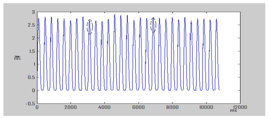
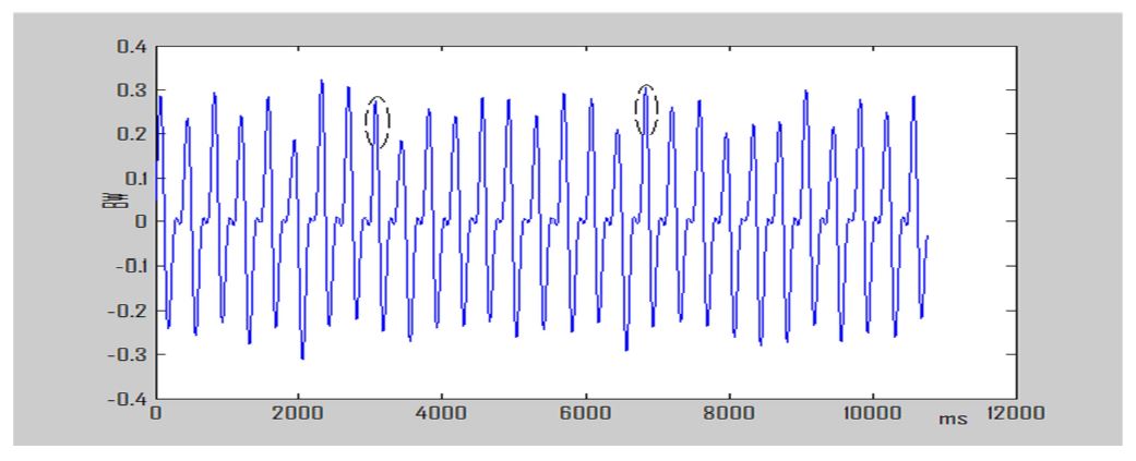
All participants performed sufficient warm-up before the experiment and wore the same type of shoes provided by the researcher (Prospecs Flash101-103). They ran at fixed speeds of 8, 12, and 16 km/h on a treadmill equipped with a force plate (instrumented dual belt tread- mills, Bertec, USA) for 3 min. The fixed speeds were determined by measuring the maximum speed that can be maintained for 3 min (16 km /h) by women who have relatively lower running abilities and decreasing the speed by 4 km/h. Prior to running at fixed speeds, the running speed was gradually increased for 30 s until the target speed was reached, and a subject ran for 30 s for a warm-up to get used to the target speed. Next, the subject ran at the fixed speed for 120 s. GRF signals were collected from 30 steps at a sampling frequency of 1,000 Hz without the subject's awareness. Data were randomly collected for the three different speed conditions. For data analysis, of the 30 steps made by the right and left feet, each, the fifth and tenth steps of the right foot were analyzed as shown in (Figure 1 and Figure 2), and their mean was obtained. Collected data were subjected to fourth-order low-pass Butterworth filtering, and the values of selected variables were obtained. The cut-off frequency was determined by calculating the power spectrum densities (PSD) of the vertical and anteroposterior GRF signals and finding the maximum frequency at which the cumulative frequency of all these signals reached 99.9% (Ryu, 2013). Ground contact was defined as a vertical GRF of 5 N or greater.
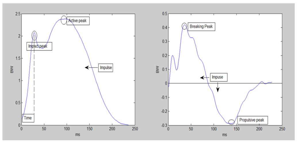
The selected variables were the impact force peak of the vertical GRF; active force peak; time to passive peak; impact loading rate, which is calculated by dividing the impact force peak by the time to passive peak; and vertical impulse, which is the integral of the vertical GRF signal with respect to a time function (Figure 3). For the anteroposterior GRF force, the anteroposterior GRF signals were integrated with respect to the breaking force peak, propulsive force peak, and time function to calculate the absolute anteroposterior impulse(Figure 3). All variables except for time are expressed in terms of weights.
3. Statistical analysis
One-way ANOVA was used to investigate the differences in the variables at different running speeds within and between genders. The level of statistical significance was set at p-value<.05.
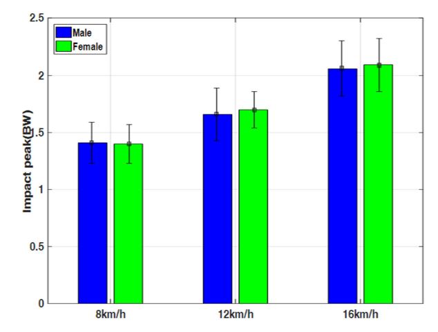
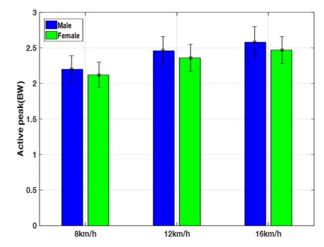
The results of the statistical analysis on the mean values and standard deviations of the variables analyzed under different speed conditions between and within genders are shown in Table 1. The impact peak that occurs at the moment the foot touches the ground while running was greater in women than in men at the relatively fast running speeds of 12 and 16 km/h, but not by a significant difference (Figure 4). The maximum active force peak per body mass, which is a vertical GRF that occurs at the moment the foot leaves the ground while running, was greater in men than in women at all running speeds, but not by a significant difference (Figure 5).
|
Ip |
Ap |
T |
Ilr |
Bp |
Pp |
Vm |
Ym |
||
|
8 km/h |
Male |
1.41±0.18 |
2.20±0.19 |
39.75±7.38 |
37.09±10.32 |
0.23±0.04 |
0.13±0.02 |
0.37±0.01 |
0.02±0.00 |
|
Female |
1.40±0.17 |
2.12±0.18 |
30.10±8.24 |
51.25±20.32 |
0.25±0.02 |
0.15±0.03 |
0.37±0.02 |
0.03±0.00 |
|
|
p-value |
.8557 |
.1971 |
.0004 |
.0085 |
.0974 |
.0255 |
.6800 |
.0359 |
|
|
12 km/h |
Male |
1.66±0.23 |
2.46±0.20 |
33.5±8.7 |
53.04±16.20 |
0.36±0.05 |
0.23±0.03 |
0.35±0.01 |
0.03±0.00 |
|
Female |
1.70±0.16 |
2.36±0.19 |
21.90±5.49 |
82.88±23.83 |
0.36±0.05 |
0.23±0.04 |
0.34±0.02 |
0.03±0.01 |
|
|
p-value |
0.4662 |
0.1047 |
0.0001 |
0.0004 |
0.9979 |
0.6608 |
0.0766 |
0.9405 |
|
|
16 km/h |
Male |
2.06±0.24 |
2.58±0.22 |
30.60±6.18 |
72.10±26.28 |
0.48±0.09 |
0.31±0.02 |
0.33±0.01 |
0.04±0.00 |
|
Female |
2.09±0.23 |
2.47±0.19 |
22.60±8.12 |
105.70±41.50 |
0.51±0.06 |
0.33±0.04 |
0.31±0.02 |
0.04±0.01 |
|
|
p-value |
0.6825 |
0.0910 |
0.0012 |
0.0040 |
0.2548 |
0.0501 |
0.0160 |
0.1572 |
|
|
8-12-16 |
Male p-value |
0.001 |
0.0001 |
0.001 |
0.0001 |
0.0001 |
0.0001 |
0.0001 |
0.0001 |
|
Female p-value |
0.0001 |
0.0001 |
0.0001 |
0.0001 |
0.0001 |
0.0001 |
0.0001 |
0.0001 |
|
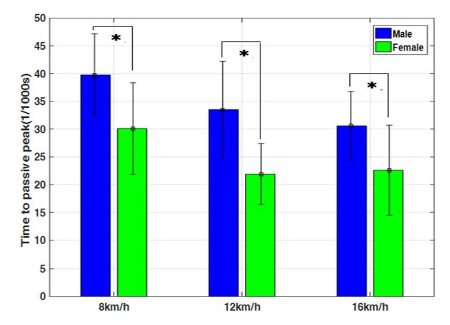
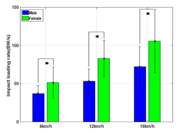
However, the mean time to maximum impact force peak was significantly shorter in women (8 km/h=30.10 ms, 12 km/h=21.90 ms, 16 km /h=22.60 ms) than men (8 km/h=39.75 ms, 12 km/h=33.5 ms, 16 km/ h=30.06 ms) at all running speeds (p<.05, Figure 6). The mean loading rate obtained by dividing the vertical impulse peak by the time to peak was significantly higher in women (8 km/h=51.25 BW/s, 12 km/h=82.88 BW/s, 16 km/h=105.70 BW/s) than men (8 km/h=37.09 BW/s, 12 km/ h=53.04 BW/s, 16 km/h=72.10 BW/s) at all running speeds (p<.05, Figure 7).
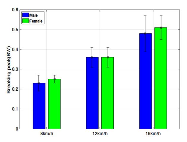
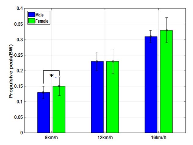
There was no significant difference in the propulsive force peak that occurred in the posterior direction when the foot came in contact with the ground between both genders at all running speeds (Figure 8). However, the mean propulsive force peak in the anterior direction was significantly greater in women (0.15 BW) than in men (0.13 BW) at the slowest running speed of 8 km/h (p<.05, Figure 9). No significant difference in the mean propulsive force peak in the anterior direction was observed for the other running speeds.
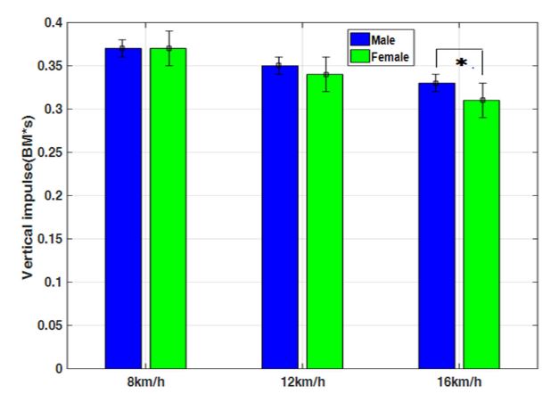
No significant difference in the vertical impulse, which is calculated by the area under the vertical GRF curve over time in the stance phase, during which the feet are in contact with the ground, was observed at 8 and 12 km/h. However, the mean vertical impulse was significantly higher in men (0.33 BW·s) than in women (0.31 BW·s) (p<.05, Figure 10).
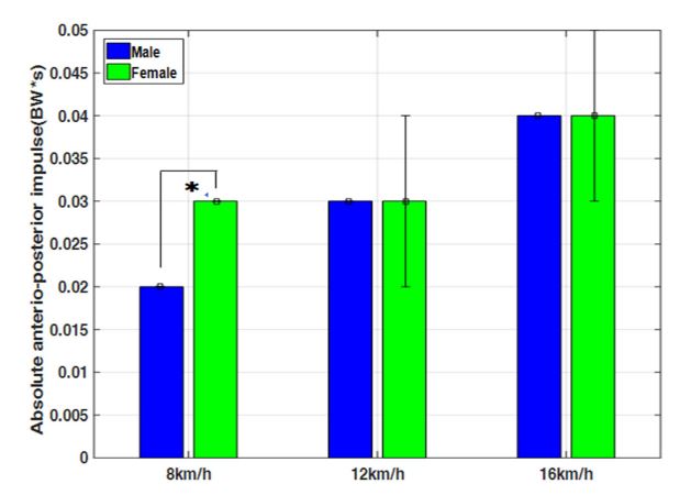
The anteroposterior impulse, which is calculated by the absolute area under the anteroposterior GRF curve in the stance phase, was significantly higher in women (0.03 BW·s) than in men (0.02 BW·s) at 8 km/h, but no significant difference was observed at the other running speeds (Figure 11).
The impact force peak, active force peak, impact loading rate, breaking force peak, propulsive force peak, and absolute anteroposterior impulse significantly increased in men and women as the running speed increased (p<.05). However, the vertical impulse significantly decreased in men and women as the running speed increased (p<.05).
This study was conducted to characterize the gender differences in GRF components, which are important determinants of running, that occur as the running speed is increased.
In this study, the time to impact force peak that occurs at the moment the foot is in contact with the ground was shorter in women than in men at all running speeds. Although a more detailed analysis on this gender difference is necessary, this finding is related to a previous report that women, who have relatively short statures, run at higher frequency rates than men at fixed running speeds and thus experience a shorter stance phase (Ferber et al., 2003). It is also possible that the time to impact force peak was shorter in women since they have shorter strides than men due to the height difference (Elliott & Blanksby, 1979). The peak value of impact force, which is known as an important factor to be studied to understand the cause of running injuries (Dorn, Schache, & Pandy, 2012), increased in proportion to the running speed in both men and women. However, no difference in the impact force peak was observed between men and women at all running speeds. The linear increase in the impact force peak with respect to the running speed was consistent with a previous finding (Dorn et al., 2012). However, considering that there was no difference in the impact force peak between both genders, it appears that gender differences do not affect impact force peaks. A comparison of foot segment angles that affect the impact force peak and vertical impulse speed between men and women is needed (Kline & Williams, 2015). However, due to the shorter time to impact force peak in women compared with that in men, the impulse loading rate per body mass was higher in women than in men. This is consistent with a previous report that female athletes have higher impulse loading rates than male athletes (Milner et al., 2006; Ryu, 2005). Previous findings have shown that the impulse loading rate is associated with plantar fasciitis (Pohl et al., 2008), patellofemoral pain, medial tibial stress syndrome, lower back pain, and hip joint and knee joint pain (Pohl et al., 2008; Hamill et al., 2008) and potential tibial stress fracture as it affects the bones that absorb early impact forces that arise during the landing phase of running (Milner et al., 2006). Therefore, the lower impulse loading rates observed in women compared with those in men at all running speeds in this study suggest that the risk of fatigue fracture is higher in women (Taunton et al., 2002), who have lower bone densities and have menstrual cycles (Bennell, Matheson, Meeuwisse, & Bruker, 1999). Female athletes may also be at increased risk of running injuries such as plantar fasciitis and lower limb pain. Since there was no difference in the impact force peak between men and women in this study, it appears that women, whose lower limbs are shorter and have less mass compared with those of men, have a disadvantage in terms of the ability of their bones to absorb impact forces while running.
Considering the significant increase in the maximum active peak as the running time was increased, it can be estimated that the damage to the muscles that absorb this force is large. However, no gender difference in the level of damage to the soleus and gastrocnemius muscles that absorb the maximum active force between the genders was observed (Kline & Williams, 2015; Schache, Dorn, Williams, Brown, & Pandy, 2014).
In this study, vertical impulse was significantly higher in men than in women at 16 km/h. Vertical impulse during running is known as an important factor that increases the level of achievement by maintaining a high step rate in situations such as muscle fatigue (Hunter, Marshall, & Cnair, 2005). Thus, it appears that the lower limb muscles play a more significant role in improving the level of achievement in male than female runners at fast running speeds (Weyand, Sternlight, Bellizzi, & Wright, 2000). In this study, aside from the time variable, a variable that significantly decreased as the running speed was increased for both men and women was vertical impulse. The reason why vertical impulse decreases as the running speed is increased may be that the leg muscles have enough time to generate a force needed to accelerate the body (Dorn et al., 2012) and create large vertical GRFs at low running speeds, whereas at relatively high running speeds, the ability of the leg muscles to generate the vertical GRFs is reduced due to the reduced time of contact with the ground (Weyand et al., 2000).
In this study, there was no difference in the maximum breaking GRF, which is created in the posterior direction in the early stance phase of running between both genders at all running speeds. However, the maximum propulsive force, which works in the anterior direction in the late stance phase, was significantly higher in women than in men at the low running speed of 8 km/h. Although detailed research on the causes of this phenomenon is necessary, a possible explanation is that female athletes extend their hip, knee, and ankle joints sufficiently at the moment their foot leaves the ground while running at low speeds (Hunter et al., 2005). It may be because of this reason that the absolute anteroposterior impulse was greater in women than in men at 8 km/h.
The significant increase in the propulsive force peak observed for both men and women as the running speed was increased is consistent with previous reports that the maximum running speed is significantly associated with the propulsive components of the anteroposterior GRF (Brughelli, Cronin, & Chaouachi, 2011; Hunter et al., 2005; Morin et al., 2012; Morin, Edouard, & Samozino, 2011; Nummela, Keränen, & Mikkelsson, 2007).
This study was aimed at characterizing the gender differences in the vertical and anteroposterior components of the GRF that occur as the running speed is increased. Considering that the impact loading rate was greater among female athletes than among male athletes, it appears that women are more exposed to the risk of potential running injuries related to the bone and ligament. It also seems that increasing the running speed may increase the risk of running injuries. Research on the association between the type of running injuries in men and women and running mechanisms may be useful in the future.
References
1. Atwater, A. E. (1990). Gender differences in distance running. In. Cavanagh PR. Biomechanics of distance running. Human Kinetics, Champaign IL.
Crossref
2. Belli, A., Kyrolainen, H. & Komi, P. V. (2002). Moment and power of lower limb joints in running. International Journal of Sport Medicine, 23, 136-141.
Crossref
Google Scholar
3. Bredeweg, S. & Buist, I. (2011). No relationship between running related injuries and kinetic variables. British Journal of Sports Medicine, 45, 310-384.
Crossref
Google Scholar
4. Brughelli, M., Cronin, J. & Chaouachi, A. (2011). Effects of running velocity on running kinetics and kinematics. The Journal of Strength & Conditioning Research, 25, 933-939.
Crossref
Google Scholar
5. Chumanov, E. S., Wall-Scheffler, C. & Heiderscheit, B. C. (2008). Gender differences in walking and running on level and inclined surfaces. Clinical Biomechanics, 23, 1260-1268.
Crossref
Google Scholar
6. Dorn, T. W., Schache, A. G. & Pandy, M. G. (2012). Muscular strategy shift in human running: dependence of running speedd on hip and ankle muscle performance. The Journal of Experimental Biology, 215, 1944-1956.
Crossref
Google Scholar
7. Elliott, B. C. & Blanksby, B. A. (1979). Optimal stride length considerations for male and female recreational runners. British Journal of Sports Medicine, 13(1), 15-18.
Crossref
Google Scholar
8. Ferber, R., Davis, I. M. & Williams, D. S., III. (2003). Gender differences in lower extremity mechanics during running. Clinical Biomechanics, 18, 350-357.
Crossref
9. Geraci, M. C. & Brown, W. (2005). Evidence-based treatment of hip and pelvic injuries in runners. Physical Medicine & Rehabilitation Clinics of North America, 16, 711-747.
Crossref
Google Scholar
10. Hamill, J., Miller, R., Noehren, B. & Davis, I. (2008). A prospective study ofiliotibial band strain in runners. Clinical Biomechanics, 23, 1018 -1025.
Crossref
Google Scholar
PubMed
11. Hunter, J. P., Marshall, R. N. & McNair, P. J. (2005). Relationships between ground reaction force impulse and kinematics of sprint-running acceleration. Journal of Applied Biomechanics, 21, 31-43.
Crossref
Google Scholar
12. Kline, P. W. & Williams, D. S., III. (2015). Effects of normal aging on lower extreminty loading and coordination during running in males and females. International Journal of Sports Physical Therapy, 10, 901-909.
Crossref
Google Scholar
13. Lopes, A. D., Hespanhol, Jr., L. C., Yeung, S. S. & Costa, L. O. (2012). What are the main running-related musculoskeletal injuries? Asystematic review. Sports Medicine, 42, 891-905.
Crossref
Google Scholar
14. Lun, V., Meeuwisse, W. H., Stergiou, P. & Stefanyshyn, D. (2004) Relation between running injury and static lower limb alignment in recreational runners. British Journal of Sports Medicine, 38, 576-580.
Crossref
Google Scholar
15. Malinzak, R. A., Colby, S. M., Kirkendall, D. T., Yu, B. & Garrett, W. E. (2001). A comparison of knee joint motion patterns between men and women in selected athletic tasks. Clinical Biomechanics, 16, 438 -445.
Crossref
Google Scholar
16. Milner, C. E., Ferber, R., Pollard, C. D., Hamill, J. & Davis, I. S. (2006). Biomechanical factors associated with tibial stress fracture in female runners. Medicine and Science in Sports and Exercise, 38(2), 323 -328.
Crossref
Google Scholar
17. Morin, J. B., Bourdin, M., Edouard, P., Peyrot, N., Samozino, P. & Lacour, J. R. (2012). Mechanical determinants of 100-m sprint running performance. European Journal of Applied Physiology, 112, 3921-3930.
Crossref
Google Scholar
18. Morin, J. B., Edouard, P. & Samozino, P. (2011). Technical ability of force application as a determinant factor of sprint performance. Medicine and Science in Sports and Exercise, 43, 1680-1688.
Crossref
Google Scholar
19. Nummela, A., Keränen, T. & Mikkelsson, L. O. (2007) Factors related to top running speed and economy. International Journal of Sports Medicine, 28, 655-661.
Crossref
Google Scholar
20. Pohl, M. B., Mullineaux, D. R., Milner, C. E., Hamill, J. & Davis, I. S. (2008). Biomechanical predictors of retrospective tibial stress fractures in runners. Journal of Biomechanics, 41, 1160-1165.
Crossref
Google Scholar
21. Robinson, R. L. & Nee, R. J. (2007). Analysis of hip strength in females seeking physical therapy treatment for unilateral patellofemoral pain syndrome. Journal of Orthopaedic & Sports Physical Therapy, 37(5), 232-238.
Crossref
Google Scholar
22. Ryu, J. S. (2005). Gender differences in the impact magnitude and its attenuation during running. Journal of Sport Biomechanics, 15(1), 91-109.
Crossref
Google Scholar
23. Ryu, J. S. (2005). Impact chock and kinematic characteristics of the lower extremity's joint during downhill running. Journal of Sport Biomechanics, 15(4), 117-129.
Crossref
Google Scholar
24. Ryu, J. S. (2013). Effects of a prolonged-run-induced fatigue on the ground reaction force components. Korean Journal of Sport Biomechanics, 23(3), 225-233.
Crossref
Google Scholar
25. Schache, A. G., Blanch, P., Rath, D., Wrigley, T. & Bennell, K. (2003). Differences between the sexes in the three-dimensional angular rotations of the lumbo-pelvic-hip complex during treadmill running. Journal of Sports Sciences, 21, 105-118.
Crossref
Google Scholar
26. Schache, A. G., Dorn, T. W., Williams, G. P., Brown, N. & Pandy, M. G. (2014). Lower-lime muscular strategies for increasing running speed. Journal of Orthopaedic & Sports Physical Therapy, 44(10), 813-824.
Crossref
27. Simoneau, G. G., Hoenig, K. J., Lepley, J. E. & Papanek, P. E. (1998). Influence of hip position and gender on active hip internal and external rotation. Journal of Orthopaedic & Sports Physical Therapy, 28, 158-164.
Crossref
Google Scholar
PubMed
28. Sinclair, J., Greenhalgh, A., Edmundson, C. J., Brooks, D. & Hobbs, S. J. (2012). Gender Differences in the Kinetics and Kinematics of Distance Running: Implications for Footwear Design. International Journal of Sports Science & Engineering, 6(2), 118-128.
Crossref
Google Scholar
29. Sinclair, J. S. & Taylor, P. J. (2014). Sex differences in tibiocalcaneal kinematics. Human Movement, 15(2), 105-109.
Crossref
Google Scholar
30. Sineclair, J., Chockalingam, N. & Vinccent, H. (2014). Gender differenced in multi-segment foot kinematics and plantar fascia strain during running. The Foot and Ankle Online Journal, 7(4), 2.
Crossref
Google Scholar
31. Taunton, J. E., Ryan, M. B., Clement, D. B., McKenzie, D. C., Lloyd-Smith, D. R. & Zumbo, B. D. (2003) A prospective study of running injuries: the Vancouver Sun Run "In Training" clinics. British Journal of Sports Medicine, 37, 239-244.
Crossref
Google Scholar
32. Taunton, J. E., Ryan, M. B., Clement, D. B., McKenzie, D. C., Lloyd-Smith, D. R. & Zumbo, B. D. (2002). A retrospective case-control analysis of 2002 running injuries. British Journal of Sports Medicine, 36, 95 -101.
Crossref
Google Scholar
33. van Gent, R. N., Siem, D., van Middelkoop, M., van Os, A. G., Bierma-Zeinstra, S. M. & Koes, B. W. (2007). Incidence and determinants of lower extremity running injuries in long distance runners: A systematic review. British Journal of Sports Medicine, 41, 469-480.
Crossref
Google Scholar
34. van Mechelen, W. (1992). Running injuries. A review of the epidemiological literature. Sports Medicine, 14, 320-335.
Crossref
Google Scholar
35. Weyand, P. G., Sternlight, D. B., Bellizzi, M. J. & Wright, S. (2000). Faster top Running speeds are achieved with greater ground forces not more rapid leg movements. Journal of Applied Physiology, 89, 1991 -1999.
Crossref
Google Scholar
36. Willson, J. D., Loss, J. R., Willy, R. W. & Meardon, S. A. (2015). Sex differences in running mechanics and patellofemoral joint kinetics following an exhaustive run. Journal of Biomechanics, 48(15), 4155 -4159.
Crossref
Google Scholar
PubMed
